Manikya Bhasma as Nanomedicine for Cancer Cells Treatment and its Characterization Using Modern Scientific Tools
Download
Abstract
Objective: This study is focused on evaluating the safety and effectiveness of Manikya Bhasma, an Ayurvedic preparation, as a nanomedicine for cancer treatment, and exploring its potential therapeutic properties using modern scientific tools.
Methods: Manikya Bhasma, composed of purified ruby, orpiment, and arsenic sulfide, was analyzed using XRD (X-ray Diffraction) for crystallite size determination, FESEM (Field Emission Scanning Electron Microscopy) for morphological analysis, EDS (Energy Dispersive X-ray spectroscopy) for elemental composition, and FTIR (Fourier Transform Infrared Spectroscopy) for functional group identification. The anticancer activity of Manikya Bhasma nanoparticles was assessed against breast cancer (MCF-7) and lung cancer (A-547) cell lines at varying concentrations (0-1000 µg/mL).
Results: The XRD analysis revealed an average crystallite size of 60 nm. FESEM micrographs confirmed the uniform distribution of particles within the sample. Notably, Manikya Bhasma demonstrated a clear dose-dependent anticancer activity against both MCF-7 and A-547 cell lines, providing reassurance about its potential effectiveness.
Conclusion: This study suggests that Manikya Bhasma has the potential to be a promising nanomedicine for cancer treatment, highlighting the therapeutic relevance of traditional Ayurvedic medicine in modern scientific research. This research serves as a bridge between traditional and modern approaches, offering new insights into the application of Ayurvedic formulations in contemporary medicine.
Introduction
Cancer is a global health concern and a leading cause of morbidity and mortality worldwide. Cancer is a major health challenge globally, affecting millions of people and causing significant societal and economic burdens [1]. The World Health Organization (WHO) estimates that cancer-related deaths will continue to increase, reaching more than 13 million deaths annually by 2030. Effective cancer treatment is crucial for reducing this burden and improving patient outcomes [2]. Ayurvedic medicine plays a vital role as a complementary and alternative approach to cancer treatment. With its holistic approach, Ayurveda addresses the physical, mental, and spiritual aspects of health. It emphasizes personalized treatments based on the individual’s constitution, lifestyle, and disease stage. Ayurvedic medicine aims to restore the balance of doshas (energetic forces) in the body and enhance overall well-being, making it a valuable addition to cancer treatment. While Ayurvedic medicine plays a role in cancer treatment, it is important to emphasize that it should be integrated with evidence-based conventional medicine [3].
Ayurveda, the ancient Indian system of medicine, offers a holistic approach to healthcare, including using Ayurvedic formulations such as Bhasma to treat various diseases, including cancer [1]. Bhasma refers to the ash or calcined form of minerals and metals, which are processed through a rigorous purification and calcination process. Ayurvedic Bhasma formulations are believed to have immune-modulating properties, which can help strengthen the body’s defense mechanisms against cancer cells. Tamra Bhasma (copper ash) [4] and Vanga Bhasma (tin ash) [5] are known for their immunomodulatory effects and may aid in enhancing the immune response to fight cancer. Mukta Bhasma (pearl ash), Praval Bhasma (coral ash), and Rajata Bhasma (silver ash) [6] are believed to possess anticancer properties and may inhibit the growth and proliferation of cancer cells. Abhraka Bhasma (mica ash) [7] and Mandura Bhasma (iron ash) are commonly used for their detoxifying and antioxidant effects. Loha Bhasma (iron ash) [8] and Makardhwaja are often used for their supportive properties for cancer treatment. Indeed, Ayurveda, an ancient medicinal system, contains a wide range of herbs with potential anticancer properties. Garlic contains organosulfur compounds, such as allicin, which possess anticancer properties. It exhibits potential in preventing and inhibiting the growth of cancer cells, particularly in breast, lung, colon, and prostate. In Ayurveda, a number of herbs, such as Green Tea (Camellia sinensis), Amalaki (Emblica officinalis), Mulethi, and Tulsi (Ocimum tenuiflorum), have anticancer activity [9].
In recent years, nanomedicine has emerged as a promising approach for the diagnosis, treatment, and prevention of various diseases, including cancer. NPs (Nanoparticles) have tremendous potential for delivering therapeutic agents directly to target cells, enhancing drug efficacy, and reducing side effects [10]. Among the wide range of nanoparticles investigated for cancer therapy, metallic nanoparticles have garnered significant attention due to their unique physicochemical properties and potential therapeutic applications [11].
Manikya Bhasma, a traditional Indian Ayurvedic medicine derived from red coral, has been extensively used in traditional medicine for its perceived medicinal properties [12]. The integration of traditional knowledge with modern scientific approaches has paved the way for exploring the therapeutic potential of Manikya Bhasma in nanomedicine for cancer treatment [9]. Recent studies have shown that the conversion of Manikya bhasma into nanoscale particles, known as Manikya bhasma nanoparticles (MBNPs), can enhance its pharmacological properties and facilitate targeted delivery to cancer cells [13]. The successful development and application of MBNPs as nanomedicines for cancer treatment require a comprehensive understanding of their physicochemical properties, drug loading capacities, and interactions with cancer cells. Characterization techniques play a crucial role in providing valuable insights into the morphology, size distribution, surface charge, crystallinity, and stability of MBNPs. Furthermore, understanding the cellular uptake and internalization mechanisms of MBNPs within cancer cells is vital for evaluating their potential efficacy and toxicity [5].
This research article aims to provide a comprehensive characterization of the Manikya Bhasma nanomedicine (MBN) and its impact on cancer cell viability by employing modern scientific tools. These techniques enable the determination of the MBNP size, shape, surface properties, crystal structure, and chemical composition [12]. The findings of this study will contribute to the current understanding of the physicochemical properties and biological interactions of MBNPs and their implications for cancer cell viability. This knowledge is essential for the rational design and development of MBN-based nanomedicine for effective cancer therapy [13]. It is also essential to conduct rigorous scientific research to further explore the efficacy, safety, and potential interactions of Ayurvedic interventions in cancer treatment. This evidence-based approach can help establish the role of Ayurvedic medicine as a valuable component of comprehensive cancer care in the modern medical landscape.
Material and characterization
Manikya Bhasma and Chemical: Manikya Bhasma was processed using standard procedures and included steps namely Manikya Shodhana (purification) and Manikya Marana (Calcination) [14]. The chemicals required for processing and testing the samples. The National Center for Cell Science in Pune, India, is where lung cancer cells (A-547) and breast cancer cells (MCF-7) cell lines were obtained [15]. Adjustable multichannel pipettes, fetal calf serum, MTT reagent, DMSO, 96-well cell culture plate (from Corning, USA), T25 flask, and 50 ml centrifuge tube were purchased from Himidiya chemicals. We purchased 5 ml centrifuge Tubes, 10 ml serological pipettes, 10 to 1000 μl pipette tips, and a Petri dish from TORSON Glassware. The sodium chloride and ethanol from Merck, Germany. Physico-Chemical Characterization: The physical characterization of a Manikya bhasma powder sample via XRD, FESEM, EDS, FTIR, and magnetic measurements [4]. FESEM is a powerful imaging technique that provides high-resolution images of the surface morphology and microstructure. The powder sample was coated with gold (40 mills/second) to make it a conductive layer to prevent charging during imaging. EDS is often combined with FESEM and provides elemental analysis of the sample. The powder sample is mixed with a suitable matrix (usually KBr) and compressed into a thin pellet to measure the FTIR spectra [16]. SQUID magnetometry was used to measure the magnetic properties, such as the magnetization and coercivity, of the samples [16]. Centrifuge (Remi), Elisa plate reader (iMark, Biorad, USA) at 540 nm and 660 nm, inverted microscope (Olympus ek2) using Camera (AmScope digital camera 10 MP Aptima CMOS) and IC50 was calculated by using software Graph Pad Prism -6.
Results
X- ray diffraction analysis
The crystallographic phase of the manikya Bhasma powder sample was analysed by X-ray diffraction technique. The XRD pattern was recorded by employing a Rigaku X-ray diffractometer (TTRX-III X-ray diffractometer, Japan) with a CuKα (1.54 Å) radiation source. The X-ray diffraction pattern of Manikya Bhasma is depicted in Figure 1.
Figure 1. X-ray Diffraction Pattern of Manikya Bhasma.
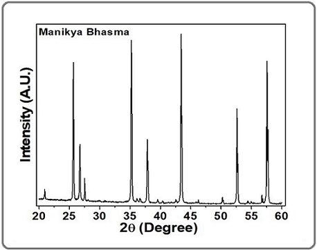
All the intense peaks in the XRD pattern of Manikya bhasma were identified and matched, and these details are given in Table 1.
| Pos. [°2Th.] | Rel. Int. [%] | Matched by Database |
| 20.957 | 6.13 | 00-046-1045 |
| 25.6564 | 81.28 | 00-010-0173 |
| 26.7223 | 31.87 | 00-033-1161; 00-046-1045 |
| 35.2229 | 97.08 | 00-010-0173 |
| 36.1304 | 1.79 | 00-038-1479 |
| 36.6317 | 2.3 | 00-033-1161; 00-046-1045 |
| 37.8374 | 37.01 | 00-010-0173 |
| 39.5412 | 2.04 | 00-033-1161; 00-046-1045 |
| 40.3628 | 1.24 | 00-033-1161; 00-046-1045 |
| 42.5295 | 1.48 | 00-033-1161; 00-046-1045 |
| 43.4185 | 100 | 00-010-0173; 00-024-0072 |
| 45.8837 | 0.98 | 00-033-1161; 00-046-1045 |
| 46.2457 | 2.11 | 00-010-0173 |
| 50.2194 | 3.9 | 00-033-1161; 00-038-1479; 00-046-1045 |
| 52.6148 | 56.59 | 00-010-0173 |
| 54.9485 | 1.22 | 00-033-1161; 00-038-1479; 00-046-1045 |
| 57.5614 | 84.92 | 00-010-0173; 00-024-0072; 00-033-0664 |
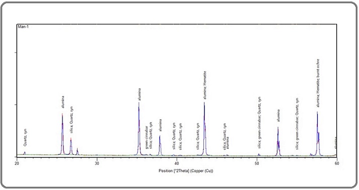
The XRD analysis found that the XRD pattern matched the different compounds available in the Manikya Bhasma with the standard ICCD database [17]. The XRD pattern of Manikya Bhasma was analysed with the help of Xpert High Score Plus software. The crystallite size of Manikya Bhasma has been estimated 60 nm by employing Scherrer formula [18]. The XRD patterns with compound names (such as silica, alumina, and iron oxide) are depicted in Figure 2.
Figure 2. X-ray Diffraction Pattern of Manikya Bhasma with Compound Names.
Microstructure and morphology analysis
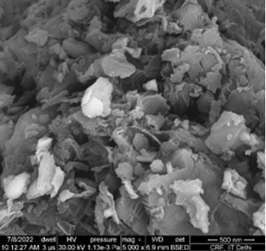
The FESEM micrographs of Manikya Bhasma are shown in Figure 3. The Manikya Bhasma is in powder form. A small amount of powder sample was dispersed onto a sample stub, which was already stuck with carbon conducting tape on the stub. The Manikya Bhasma is nonconducting, a thin conductive coating of gold was applied on the sample to prevent charging effects during the analysis [19]. The coating can also improve the image quality. The individual particles’ morphology, shape, and particle size distribution were studied [9].
Figure 3 shows that the particle size distribution and morphology are homogeneous in nature.
Figure 3. FESEM micrograph of the Manikya Bhasma.
The average particle size distribution is demonstrated through the histogram in Figure 4, and the distribution of particles ranges from 50 nm to 350 nm.
Figure 4. Histogram of the Particle Size Distribution of Manikya Bhasma.
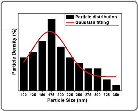
The average particle size is approximately 175 nm, as shown in the histogram, but also indicates the presence of agglomeration, which means that the individual particles in the sample tend to cluster or stick together, forming larger agglomerates [20]. Agglomeration is a common phenomenon, especially in powder samples, and can significantly affect the material’s properties and applications [18].
Energy dispersive X-ray spectroscopy (EDS) analysis
Energy dispersive X-ray spectroscopy is often combined with field emission scanning electron microscopy (FESEM) to analyse the elemental composition of a sample. In this technique, the collected X-rays are sorted based on their energies by an EDS detector, for the Manikya bhasma.
The weight percentages and atomic percentages of the elements obtained from Manikya Bhasma are shown in Table 2.
| Element | Weight% | Atomic% |
| C K | 53.93 | 63.77 |
| O K | 36.21 | 32.14 |
| Na K | 0.58 | 0.36 |
| Mg K | 0.61 | 0.36 |
| S K | 2.71 | 1.2 |
| Cl K | 0.9 | 0.36 |
| K K | 2.2 | 0.8 |
| Ca K | 2.86 | 1.01 |
| Totals | 100 |
This Bhasma contained carbon, oxygen, sodium, magnesium, silicon, chlorine, potassium, and calcium. EDS analysis is a valuable tool in pharmatutical industries [20]. The characterization of Manikya bhasma and determining its elemental compositions. Each element produces characteristic peak in the spectrum, allowing for qualitative elemental identification. This information is very important in the medical field of ancient Aurvedic Bhasma for proper doage and proper application [13]. The chemical composition of the Ayurvedic Bhasma is crucial for verifying its quality and ensuring that it meets the specifications required for safe use in Ayurvedic practices.
Fourier transform infrared (FTIR) spectroscopy
FTIR spectroscopy is a nondestructive and powerful analytical tool for the qualitative and semiquantitative analysis of Manikya bhasma and other Ayurvedic medicines [7]. It can provide valuable information about the chemical composition, functional groups, and purity of the medicine, helping to ensure its safety and efficacy for medicinal use.
FTIR spectra are obtained in the mid-infrared range, typically ranging from 400 cm-1 to 4000 cm-1. This range covers a wide variety of functional groups and molecular vibrations, making FTIR a powerful technique for identifying and characterizing organic and inorganic compounds. FTIR spectra contain absorption peaks at specific wavenumbers, each corresponding to different functional groups present in the sample [21]. For Manikya bhasma, absorption peaks in the range of 400 to 4000 cm-1 are depicted in Figure 5 (a), and absorption peaks in the range of 400 cm-1 to 4000 cm-1 are clearly visible in Figure 5 (a) and (b).
Figure 5. Fourier Transform Infrared Spectroscopy Spectra of Manikya Bhasma.
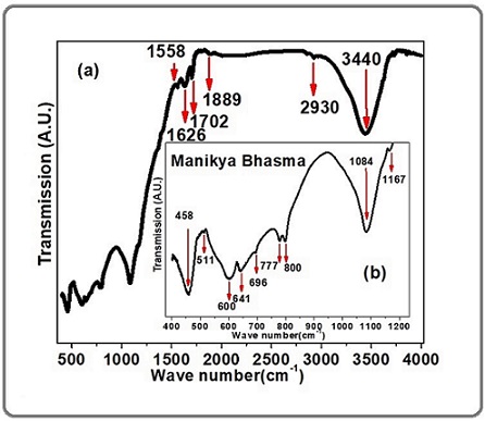
These absorption peaks show information about the presence of various organic and inorganic functional groups of Manikya Bhasma.
The vibrational modes present in Minkya Bhasma are listed in Table 3.
| Wavenumber (cm-1) | Appearance | Compound Name | Vibrational modes |
| 458 | Strong | Metal-Oxide | |
| 511 | Medium | Mg-O | |
| 600 | Medium | Ca-O | |
| 641 | Medium | Si-O | |
| 696 | Medium | Ca-O | |
| 777 | Medium | C-Cl | |
| 800 | Medium | alkene | C-C bending |
| 1084 | Strong | Si-O-Si | |
| 1167 | Medium | ether | C-O stretching |
| 1558 | Medium | amine | H-O-H |
| 1626 | Medium | ketone | C=C stretching |
| 1702 | Medium | C=O stretch | |
| 1889 | Strong | aromatic compound | C-H bending |
| 2930 | Medium | Alkanes | C-H stretch |
| 3440 | Strong | Alcohol | O–H stretch |
Fifteen vibrational modes were observed in the 400 cm-1 to 4000 cm-1 FTIR spectra of Manikya Bhasma. Among the 15 absorption peaks, some are strong, whereas some are weak. The vibrational modes present in the range of 458 to 700 cm-1 wavenumbers for metal oxide bonds such as Mg-O, Ca-O, and Si-O. A strong absorption peak is observed at 1084 for Si-O-Si vibrational modes. A strong absorption peak was observed at 3440 cm-1, which represents the alcohol groups of compounds with O-H stretching vibrations [13]. These observed vibrational modes of metal oxides are also supported by the XRD analysis in the previous section of this article and the EDS elemental analysis.
Magnetic Properties
Magnetic characterization of Ayurvedic bhasma (nanomedicine) involves the study of magnetic properties at the nanoscale, and these properties are exploited for various biomedical applications, including targeted drug delivery, hyperthermia, and magnetic resonance imaging (MRI). Magnetic characterization is essential for understanding and optimizing the performance of these nanomaterials for specific medical applications [4], [22].
The magnetic properties (M-H loops) of the Manikya bhasma are shown in Figure 6.
Figure 6. Magnetic Property (M-H loop) of Manikya Bhasma.
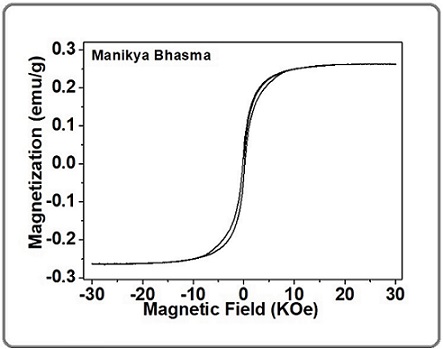
It is characterized by employing superconducting quantum interference device (SQUID) magnetometry. A very low magnetization of 0.3 emu per gram was observed. Understanding the magnetic properties of ayurvedic nanomedicine to determine the biocompatibility and potential toxicity of magnetic nanoparticles is essential for safe medical applications [16]. Cell viability assays and in vivo studies were conducted to evaluate the cytotoxicity and biocompatibility of the nanoparticles.
Manikya Bhasma has nanomedicine-like characteristics
The features of nano ayurvedic Bhasma, such as crystal size, surface morphology, presence of elements, presence of functional groups, and other factors, determine its nano medicinal impact [9]. The morphology of Manikiya Bhasma nanoparticles, which range in size from 50-300 nm with an average of 175 nm, is depicted in the FE-SEM micrograph of Manikya Bhasma (Figures 3). The XRD analysis estimated the average crystallite size of Minikiya Bhasma was 60 nm. The nanoparticles of Minikiya Bhasma has been analysed with help of FTIR, which estimated the present functional groups. EDS technique has estimated the present elements of Minikiya Bhasma [13]. This meant that the size may be linked to the particle’s biological activity, meaning that Manikiya Bhasma has favorable features as a nanomedicine for cancer treatments.
Manikya Bhasma induces cytotoxicity in cancer cells
Cell culture: The cytotoxicity test of breast cancer cell (MCF-7) and lung cancer cells (A-547) (Procured from NCCS Pune) cell line was determined using an MTT Assay. Cells (10,000 cells/well) were cultured in 96 well plates for 24 h in DMEM (Dulbecco’s Modified Eagle Medium) [23]. It was supplemented with 10% Fetal bovine serum (FBS) and 1% antibiotic solution at 37°C with 5% CO2. After the 24 hours, the cells were treated from (as per mention in the excel sheet) of the formulations (different concentrations were prepared in incomplete medium) [24]. Cells without treatment were considered as Control. After incubation for 24 hours, MTT Solution (a final concentration of 250µg/ml) was added to cell culture and further incubated for 2 h. At the end of the experiment, culture supernatant was removed and cell layer matrix was dissolved in 100 µl Dimethyl Sulfoxide (DMSO) and read in an Elisa plate reader (iMark, Biorad, USA) at 540 nm and 660 nm. IC50 was calculated by using software Graph Pad Prism -6. Images were captured under inverted microscope (Olympus ek2) using Camera (AmScope digital camera 10 MP Aptima CMOS) [25].
% cell viability=(Abs of treated cells)/(Abs of Untreated cells) x100
The IC50 value was determined by using a linear regression equation i.e. Y =Mx+C. Here, Y = 50, M, and C values were derived from the viability graph [26].
Cytotoxicity in cancer cells
Manikya Bhasma is an herbal medicine that alters cell metabolism, preventing the growth and metastasis of cancer cells. Determining the role of Manikya Bhasma as an anti-inflammatory nanomedicine [13]. Breast cancer cells (MCF-7) and lung cancer cells (A-547) were treated with Manikya Bhasma (0-1000 µg/mL) for 48 h, and cell viability was determined using MTT reduction assay as described in the Materials and Methods section, and Cell Culture section. The plot of cell viability (%) versus the concentration (0, 1, 10, 50, 100, 250, 500 and 1000 μg/mL) of Manikya Bhasma for breast cancer cell (MCF-7) has been shown in Figure 7.
Figure 7. Tumor Cell Viability Analysis Via MTT Assay of MCF-7 Through Manikya Bhasma.
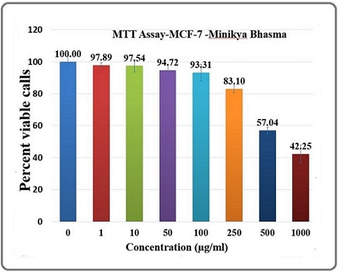
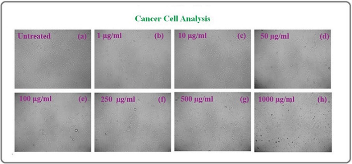
Based on the above plot of cell viability (%) versus the concentration, it was used to calculate the IC50 [27]. The Manikya Bhasma reduced the viability of breast cancer cell (MCF-7) cells in a dose-dependent fashion, with an IC50 of 804.7±0.043 μg/mL [9]. The morphology of untreated to treated effectiveness of Manikya bhasma on breast cancer cell (MCF-7) are shown in Figure 8 (a-h) for 0, 1, 10, 50, 100, 250, 500 and 1000 μg/mL samples. The Manikya Bhasma is very effective against other cancer cell line lung cancer cells (A-547).
Figure 8. Morphology of Tumor Cell Viability Analysis Via MTT Assay of MCF-7 Through Manikya Bhasma with Different Concentrations.
The Figure 9 shows that, plot of cell viability (%) versus the concentration (0, 1, 10, 50, 100, 250, 500 and 1000 μg/mL ) of Manikya Bhasma for lung cancer cells (A-547) [28].
Figure 9. Tumor Cell Viability Analysis Via MTT Assay of (A-547) Through Manikya Bhasma.
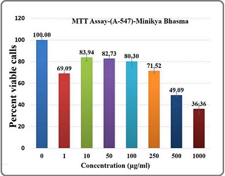
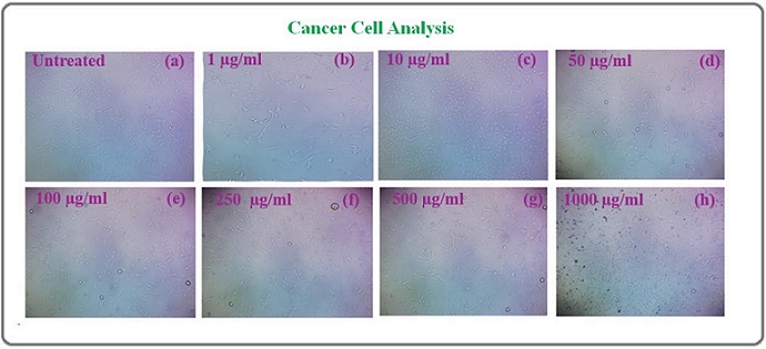
The Manikya Bhasma reduced the viability of lung cancer cells (A-547) cells in a dose-dependent fashion, with an IC50 of 736.7±0.043 μg/mL [29]. The morphology of untreated to treated celline of dose-dependent Manikya Bhasma (0, 1, 10, 50, 100, 250, 500 and 1000 μg/mL ) are shown in Figure 10.
Figure 10. Morphology of Tumor Cell Viability Analysis Via MTT Assay of (A-547) Through Manikya Bhasma with Different Concentrations.
Discussion
Nanotechnology is being studied for cancer therapeutics since nanoparticle play a major role in targeted drug delivery [30]. Nanoparticle-based drug delivery has specific advantages such as biocompatibility, stability, enhanced permeability and retention effect, and precise targeting [31]. In Indian system of medicine called Ayurveda, Bhasma is incinerated metal-mineral ash and it is having size in nanometers [32]. These Bhasma are being used successfully in clinical practice from many years. The motive behind the study was to find the anticancer activity i.e. cytotoxic potential of Manikya Bhasma using MTT assay [25].
In this article, we have aim to the study of Manikya Bhasma particle size is in nano range (~100 nm) and it potential use as nanomedicine for anticancer activity [14]. The crystallite size of Manikya Bhasma has been estimated approx. 60 nm by employing Scherrer formula through XRD patterns. It is also found that the silica, alumina, and iron oxide compounds are present in Manikya Bhasma. From the FESEM analysis, it is also found that the morphology of Manikya Bhasma is spherical in shape and the distribution of particles ranges from 50 nm to 350 nm with the average particle size is approximately 175 nm. The FTIR analysis show information about the presence of various organic and inorganic functional groups of Manikya Bhasma [33]. The elements present in the Manikya Bhasma through EDS analysis are carbon, oxygen, sodium, magnesium, silicon, chlorine, potassium, and calcium. The nano form of Manikya Bhasma was found more effective than bulk form. It shows decreasing cellular viability of cancer cell lines in a different concentration of nanomedicine [15]. The cytotoxic effects of Manikya Bhasma on non-cancer cell lines show that it is non-toxic to normal cells. The size of Manikya Bhasma is in the nanometer range, known as nanomedicine, and its effects on cancer cells.
In conclusion, the ayurvedic nanocrystalline Manikya Bhasma has been characterized by modern scientific tools such as XRD, FESEM, EDS, FTIR, and cancer cell line experiments. XRD analysis estimated that the crystallite size is 60 nm and crystalline in nature. The morphology and particle size of Manikya Bhasma have been studied by FESEM and agglomeration of the nanocrystallites with an average particle size of 175 nm. These findings may create a link between modern science and technology, as well as ancient medicine. The current study’s findings encourage the application of old Indian wisdom from Ayurveda to generate newer medications for use in modern nanomedicine. Manikya Bhasma nanoparticles were tested for their anticancer activity against breast cancer cells (MCF-7) and lung cancer cells (A-547) at various concentrations (0-1000 µg/mL). The size of Manikya bhasma is in the nanometre range, which is known as nanomedicine, and it affects cancer cells. This technique has substantial advantages for cancer treatment and side effect reduction.
Acknowledgments
The authors extend sincere gratitude and acknowledge IIT Delhi for providing access to their experimental facility available at the Central Research Facility (C. R. F.) and Dr. Rakesh Kr Singh, Head of the Aryabhatta Center for Nanoscience and Nanotechnology at Aryabhatta Knowledge University, Patna, for his valuable discussions and insights. I would like to extend my sincere gratitude to Aakaar Biotechnologies Private Limited, for Cytotoxicity Evaluation of the samples.
Financial support and sponsorship
Nil.
Author Contribution Statement
Reena: Investigation, Analysis, writing original draft. Preeti Sharma: Supervision, review and editing original draft. Nakuleshwar Dut Jasuja: Supervision, review and editing original draft. Sunil Kumar: Analysis, review and editing original draft.
Conflicts of interest
There are no conflicts of interest.
Data Availability
The authors declared that data is available within the manuscript.
Declarations
This declaration is not applicable.
References
- Integrating ayurvedic medicine into cancer research programs part 1: Ayurveda background and applications Arnold JT . Journal of Ayurveda and Integrative Medicine.2023;14(2). CrossRef
- A fusion decision system to identify and grade malnutrition in cancer patients: Machine learning reveals feasible workflow from representative real-world data Yin L, Song C, Cui J, Lin X, Li N, Fan Y, Zhang L, et al . Clinical Nutrition (Edinburgh, Scotland).2021;40(8). CrossRef
- Integrating ayurvedic medicine into cancer research programs part 2: Ayurvedic herbs and research opportunities Arnold JT . Journal of Ayurveda and Integrative Medicine.2023;14(2). CrossRef
- Study on physical properties of Ayurvedic nanocrystalline Tamra Bhasma by employing modern scientific tools Singh RK , Kumar S, Aman AK , Karim S. M., Kumar S, Kar M. Journal of Ayurveda and Integrative Medicine.2019;10(2). CrossRef
- Understanding mechanistic aspects and therapeutic potential of natural substances as anticancer agents | Request PDF Deep A, Kumar D, Bansal N, Narasimhan B, Marwaha RK , Sharma PC . ResearchGate.2024;3(2):100418. CrossRef
- Swarna Bhasma in cancer: A prospective clinical study Das S, Das MC , Paul R. Ayu.2012;33(3). CrossRef
- Study on physical properties of Indian based ayurvedic medicine: Abhrakh bhasma as nanomaterials by employing modern scientific tools Singh RK , Kumar S, Aman AK , Kumar S, Kar M. GSC Biological and Pharmaceutical Sciences.2018;5(2). CrossRef
- Study of structural, optical, and toxicity of iron-based nano particle Kasis bhasma | Request PDF Kr Diwedi P, Kr. Singh R, Kour P, Kumar N, Kumar P, Kar M. ResearchGate.2024;62:4006-4012. CrossRef
- Herbal Nanoparticles: A Commitment towards Contemporary Approach Mansingh PP . Indian Journal of Pharmaceutical Education and Research.2023;57(3s). CrossRef
- Preparation of superfine cinnamon bark nanocrystalline powder using high energy ball mill and estimation of structural and antioxidant properties Abhay AK , Rakesh SK , Nishant K, Birendra P. IOP Conference Series: Materials Science and Engineering.2021;1126(1). CrossRef
- Impact of B-Glucan Against Ehrlich Ascites Carcinoma Induced Renal Toxicity in Mice Hasan AF , Alankooshi AA , Abbood AS , Dulimi AG , Al-Khuzaay HM , Elsaedy EA , Tousson E. OnLine Journal of Biological Sciences.2023;23(1). CrossRef
- Exploring the Anticancer Potential of Traditional Thai Medicinal Plants: A Focus on Dracaena loureiri and Its Effects on Non-Small-Cell Lung Cancer Huang X, Arjsri P, Srisawad K, Yodkeeree S, Dejkriengkraikul P. Plants (Basel, Switzerland).2024;13(2). CrossRef
- Manikya Bhasma is a nanomedicine to affect cancer cell viability through induction of apoptosis Jha S, Trivedi V. Journal of Ayurveda and Integrative Medicine.2021;12(2). CrossRef
- Cytotoxic Effect of Phoenix dactylifera (Iraqi Date) Leaves and Fruits Extracts against Breast Cancers Cell Lines Al-Zeiny SSM , Mohammad MH , Almzaien AK , Al-Shammari AM , Ahmed AA , Shaker HK . Asian Pacific journal of cancer prevention: APJCP.2024;25(7). CrossRef
- Cytotoxic Activity of Hypericum triquetrifolium Turra Methanolic Extract Against Cancer Cell Lines Al-Anee RS , Al-Ani EH , Mousa ZS . Asian Pacific journal of cancer prevention: APJCP.2023;24(10). CrossRef
- Effect of lattice strain on structural and magnetic properties of Ca substituted barium hexaferrite Kumar S, Supriya S, Pandey R, Pradhan LK , Singh RK , Kar M. Journal of Magnetism and Magnetic Materials.2018;458. CrossRef
- Effect of Sintering Temperature on Electrical Properties of BHF Ceramics Prepared by Modified Sol-Gel Method Kumar S, Supriya S, Kar M. Mater Today Proc.2017;4(4):5517-5524. CrossRef
- Grain Size Effect on Magnetic and Dielectric Properties of Barium Hexaferrite (BHF) | Request PDF Kumar S, Supriya S, Pradhan LK , Pandey R, Kar M. ResearchGate.2024;579:411908. CrossRef
- Curcumin and colorectal cancer: An update and current perspective on this natural medicine Weng W, Goel A. Seminars in Cancer Biology.2022;80. CrossRef
- Effect of Microstructure on Electrical Properties of Li and Cr Substituted Nickel Oxide Kumar S, Supriya S, Pradhan LK , Kar M. J Mater Sci Mater Electron.2017;28(22):16679-16688. CrossRef
- Pharmacology, Toxicology, and Therapeutic Effects of Metals and Minerals Used in Traditional Medicine Perera PK , Dahanayake JM , Diddeniya JID . In Medical Geology; Prasad, M. N. V., Vithanage, M., Eds.; Wiley.2023;:303-313. CrossRef
- Acute and subchronic toxicity study of Tamra Bhasma (incinerated copper) prepared with and without Amritikarana Chaudhari SY , Nariya MB , Galib R., Prajapati PK . Journal of Ayurveda and Integrative Medicine.2016;7(1). CrossRef
- In vitro cytotoxicity assays: comparison of LDH, neutral red, MTT and protein assay in hepatoma cell lines following exposure to cadmium chloride Fotakis G, Timbrell JA . Toxicology Letters.2006;160(2). CrossRef
- Investigation of in-vitro Anti-Cancer and Apoptotic Potential of Garlic-Derived Nanovesicles against Prostate and Cervical Cancer Cell Lines Sharma V, Sinha ES , Singh J. Asian Pacific journal of cancer prevention: APJCP.2024;25(2). CrossRef
- Tetrazolium (MTT) assay for cellular viability and activity Morgan D. M.. Methods in Molecular Biology (Clifton, N.J.).1998;79. CrossRef
- Cell sensitivity assays: the MTT assay Meerloo J, Kaspers GJL , Cloos J. Methods in Molecular Biology (Clifton, N.J.).2011;731. CrossRef
- Tumor Educated Platelets as a Biomarker for Diagnosis of Lung cancer: A Systematic Review Walke V, Das S, Mittal A, Agrawal A. Asian Pacific journal of cancer prevention: APJCP.2024;25(6). CrossRef
- Evaluation of the Anti-Carcinogenic Effect of Centella Asiatica on Oral Cancer Cell Line: In vitro Study Vijayakumar T, Rameshkumar A, Krishnan R, Bose D, V V, G N. Asian Pacific journal of cancer prevention: APJCP.2023;24(5). CrossRef
- Anticancer Activity of Aaptos suberitoides on Glioblastoma Multiforme Cell Line Huda F, Hermawan R, Putri T, Dwiwina RG , Berbudi A, Bashari MH . Asian Pacific journal of cancer prevention: APJCP.2024;25(5). CrossRef
- Plant-derived bioactive compounds in colon cancer treatment: An updated review Esmeeta A, Adhikary S, Dharshnaa V., Swarnamughi P., Ummul Maqsummiya Z., Banerjee A, Pathak S, Duttaroy AK . Biomedicine & Pharmacotherapy = Biomedecine & Pharmacotherapie.2022;153. CrossRef
- Role of Salvia Hispanica Seeds Extract on Ehrlich Ascites Model Induced Liver Damage in Female Mice Hasan AF , Jasim N, Suhail M, Abid A, Tousson E. J Biosci Appl Res.2024. CrossRef
- Recent Advances in Elucidating the Biological Properties of Withania Somnifera and Its Potential Role in Health Benefits Alam N, Hossain M, Khalil, Md I, Moniruzzaman M, Sulaiman SA , Gan SH . Phytochem Rev.2012;11(1):97-112. CrossRef
- Natural Products as Anticancer Agents Varghese R, Dalvi YB . Current Drug Targets.2021;22(11). CrossRef
License

This work is licensed under a Creative Commons Attribution-NonCommercial 4.0 International License.
Copyright
© Asian Pacific Journal of Environment and Cancer , 2024
Author Details
How to Cite
- Abstract viewed - 0 times
- PDF (FULL TEXT) downloaded - 0 times
- XML downloaded - 0 times