Surgical Tactics for Intestinal Duplication Cyste Causing Intestinal Invagination (Case From Practice)
Download
Abstract
We give a case study of how we encountered a very rare pathology - an intestinal duplication cyst of the terminal ileum, which manifested itself very early in a child of two months with a complication of invaginal intestinal obstruction. Taking the very serious condition of the newborn into consideration, the tactics of minimally invasive surgery were used - mini laporatomy, intestinal disinvagination, excision of enterocysts, and the application of cecostomy. Combined anesthesia: Total intravenous anesthesia (sibazon + ketamine) with the preservation of spontaneous breathing + spinal anesthesia with bupivacaine 1.0 ml at the level of Th 11. This managed to avoid a more traumatic abdominal operation, thereby not only saving life, but also preventing various possible complications.
Introduction
Intestinal cysts are rare. In the literature, terminology is currently most often used to designate cysts developing along the intestinal tube - intestinal duplication cysts [1, 2]. Intestinal duplication cysts usually appear during the first years of life as a palpable mass, as well as signs of intestinal obstruction. Intestinal intussusception is the most common type of acquired intestinal obstruction in children, while in the vast majority this pathology occurs in infants (according to the literature, more often from 4 months to 2 years of age). In infants, this pathology develops against the background of anatomical and physiological features, which include the mobility of the ileum and cecum, the immaturity of the Bauhinia valve. It is precisely with these features that intussusception in children under one year old most often develops in the area of the ileocecal angle [3]. Various organic causes also lead to intussusception of the intestine: intestinal polyps, tumors, duplication of various parts of the intestine, Meckel’s diverticulum, etc. [4]. According to the literature, there is information that enterocystoma was also organic causes of intestinal intussusception, but in very rare cases. As an example, we give our own observations.
Child K.K. date of birth Octover 30,2019 (Karakalpakstan, Nukus), birth weight 4800 grams. First child. Breastfed. On the 44th day of life, he entered the Chimbay regional medical association, intensive care unit with a diagnosis of intestinal obstruction, intestinal paresis. Complaints at admission to frequent vomiting, bloating, anxiety, weakness, scanty stools, refusal to eat. From the anamnesis after birth, the child had frequent bloating, accompanied by regurgitation of gastric contents. Stool in the first month of life was normal, in the second month it became irregular, scarce.
Upon admission to the hospital, the child’s condition was extremely serious. Fecal vomiting, severe intestinal paresis, and exicosis were noted. The skin is pale and dry. Tachycardia, shortness of breath due to abdominal distention. There was no stool. At the moment of examination, the child is weakened, reacts to contact with weak anxiety, groans. Excretion of intestinal contents with an admixture of bile from the gastric tube. The abdomen is sharply distended, asymmetric in the upper abdomen, there is a weakened peristalsis of the dilated loops of the small intestine. Percussion amplification of sound in the upper sections with dullness of percussion sound in the lower abdomen, intestinal motility is sharply weakened. The abdomen is tense, the peritoneal symptoms are positive. Urine - oliguria, concentrated. On the roentgenogram, there is an expansion of the loops of the small intestine, with darkening in the lower abdomen. Ultrasound of the abdominal cavity pneumatosis of the intestine.
A team of pediatric surgeons and an anesthesiologist were summoned to the District Hospital through the sanitary aviation (on the second day after hospitalization). Considering the severity of the child’s condition (he is not transportable, the distance of the trip to the emergency medicine center is more than 60 km, the night time, the duration of the ride would take one and a half to two hours), it was decided to operate in a regional hospital with a diagnosis of intestinal obstruction complicated by peritonitis. Anesthetic risk according to ASA 1E, there is no pediatric ventilator for large cavitary surgery.
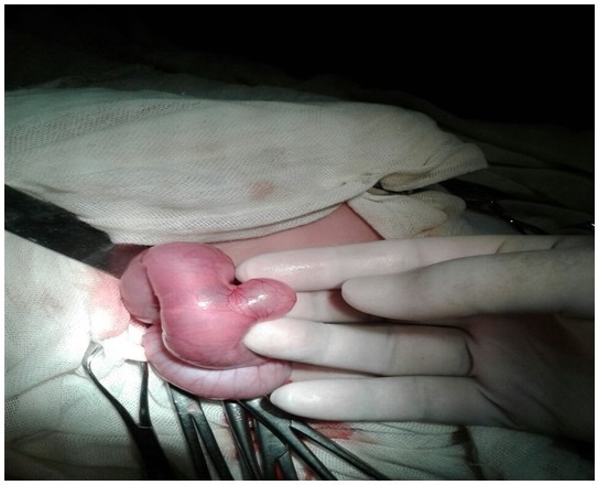
There was a question about the access of the operation, the scope of the operation, the type of anesthesia for this patient. Since there was a subjective clinic of small bowel obstruction, we chose the access - mini laparotomy according to Dyakonov-Volkovich. Combined anesthesia: total intravenous anesthesia (sibazon + ketamine) against the background of spontaneous breathing + spinal anesthesia with bupivacaine 1.0 ml at the Th 11. When the abdomen was opened, serous-purulent fluid was released from the peritoneum, and small-colonic intussusception was noted. The ileal loops are swollen. Disinvagination was performed, it turned out that the cause of intestinal intussusception was a congenital enterocystoma of the terminal part of the ileum (Figure 1).
Figure 1. Enterocyst Located in the Distal Ileum.

Enterocyst, 3.0x2.5 cm in size, partially invaded the intestine causing partial intestinal obstruction, part of the cyst protruded beyond the intestinal wall. With a diagnostic puncture, the contents are viscous, cloudy, mucous in nature. Performed marginal resection of the cyst with treatment of the cavity with antiseptic solutions. The abdominal cavity was sanitized with a solution of decasan diluted with saline, the pelvis was drained. For the purpose of decompression of the intestine, a cecostomy was imposed. Parenteral nutrition, infusion therapy, transfusion of blood components, antibiotic therapy were carried out, the child was connected to humidified oxygen. The child is not intubated for mechanical ventilation. The stomach was washed daily, hourly analgesic therapy was carried out for 5 days. Feeding began on the third day, fractional, with breast milk. Intestinal stimulation, stool was on the day after surgery after enema, intestinal stoma is functioning. The drainage tube was removed on day 3. Abdominal ultrasound in dynamics - without pathological signs.
In the dynamics in the postoperative period, the child’s condition gradually stabilized, the postoperative wound healed by secondary intention. On the 10th day, the child was discharged for outpatient observation in a relatively satisfactory condition.
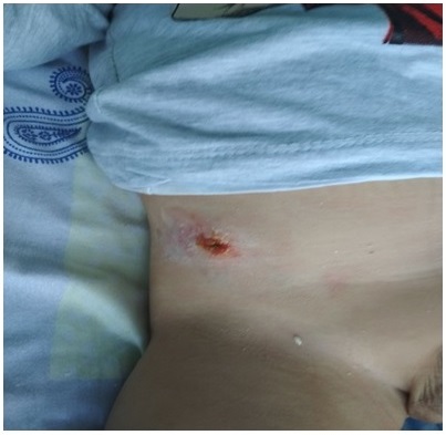
The results of the second examination after 10 months: the child’s condition is satisfactory, the age is not lagging behind, the feeding is mixed, there is no bowel paresis, the stool is independent, the cecostomy functions as a small intestinal fistula (Figure 2), the child is active, gaining weight.
Figure 2. The State of the Postoperative Wound after 10 Months, the Presence of an Intestinal Fistula.

In conclusions, our case shows that in the surgical treatment of rare forms of pathology in children (enterocyst of the terminal ileum), in limited rural conditions, the optimal choice of tactics would be the use of minimally invasive surgery under spinal anesthesia. This approach is less traumatic than abdominal surgery, it reduces the risk of various possible postoperative complications (early adhesive intestinal obstruction, ventral hernia, etc.) and deaths in children.
References
- Kishechnaia duplikacionnaiakista: uitrazvukovaia diagnostika I morfologicheskie sostoiania. // SonoAce Ultrasound . Journal - Moscow. 2010;(21): With. 64-71. (in Russian) Stepanova Y, Dubova E. .
- Completely isolated enteric duplication cyst: case report Kim S. K., Lim H. K., Lee S. J., Park C. K.. Abdominal Imaging.2003;28(1). CrossRef
- Nash opit diagnostiki I lechenia invaginacii kishechnika u detey. // Sovremennie problemi nauki i obrazovania. Moscow, 2018 No. 2. (in Russian) Barskaya M.A. and coauthors . .
- Anatomicheskie prichini kishechnoy invaginacii u detey. // Vestnik problem biologii I medicine. Moscow. 2019;1, (in Russian)(2) Gritsenko E, Gritsenko E. .
Author Details
How to Cite
- Abstract viewed - 0 times
- PDF (FULL TEXT) downloaded - 0 times
- XML downloaded - 0 times