Papillary Endometrial Adenocarcinoma with Cervical Dysplasia: Report of a Case and Review of Literature
Download
Abstract
Objective: Multifocality in gynecologic malignancies is a common phenomenon, however synchronous tumors may occur. Synchronous cancers are about 1.7% of gynecologic malignancies.
Methods: A 57-year old female with chief complaint of vaginal bleeding was admitted. Endometrial curettage and cervical biopsy was done.
Result: Pathologist reported: compatible with papillary adenocarcinoma, Grade II in endometrial sample and squamous epithelium with moderate dysplasia and tiny fragments of atypical glandular epithelium in endocervical samples. The patient refused for surgical excision of the lesion and insisted on to treat with conventional herbal medicine. Later Pap smear was done and pathologist reported: “High grade squamous intraepithelial lesion (HSIL) and atypical glandular cells, favor neoplastic in atrophic background”.
Conclusion: In the case of gynecologic cancer be careful that it may accompany another gynecologic malignancy or premalignant lesion. The second lesion may occur synchronous or metachronous or may be metastatic. Many of the synchronous malignancies are presented in lower stages and have better prognosis than metastatic lesion. Thorough sampling and examination is important in correct diagnosis and treatment.
Introduction
Multifocality in gynecologic malignancies is a common phenomenon, however synchronous tumors may occur [1]. Synchronous cancers are about 1.7% of gynecologic malignancies [2]. Most of the cases are in low stage and low grade [2-4]. More favorable prognosis is expected in synchronous cancers than metastatic or primary advanced tumors [4]. Estrogens are known as etiologic agents for endometrial and ovarian neoplasms. Human papilloma virus is considered as an etiological agent in cervicovaginal malignancies [2]. Immunohistochemistry may be necessary for differentiation between various types of gynecologic cancers [1, 5]. Here we report synchronous presentation of papillary adenocarcinoma of endometrium with dysplasia in cervix uteri in a postmenopausal lady.
Case report
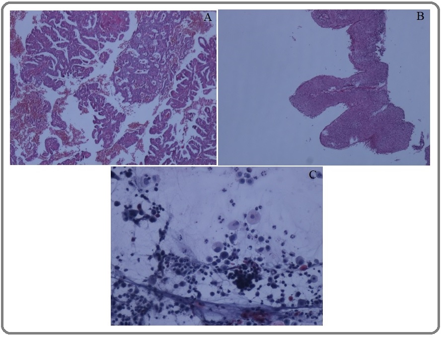
A 57-year old Gravid2, Parity2, Live child2 (G2 T2 L2, Normal vaginal delivery x II) female was referred to obstetrics and gynecologic ward with chief complaint of vaginal bleeding on April 8th 2017.She was menopause since 10 years ago. She was a teacher and had past medical history of anemia and surgery of cyst in uterine cervix. Liquid –based Papanicolaou smear (Pap smear) on March 1th 2017 report was “Atypical squamous cells cannot exclude HSIL (ASC-H), shift in flora suggestive of bacterial vaginosis and severe inflammation with recommendation for colposcopic biopsy.” Tumor markers on March 4th 2017 were normal. Ultrasound examination on March 13th 2017 demonstrated uterus measuring 63*61 mm larger than normal. Uterine cavity was contained abundant fluid with multiple polypoid areas related to endometrium with maximum dimensions of 28*14 mm. Ovaries were atrophic. Otherwise kidneys and bladder were normal. Endometrial dilatation & curettage ( D&C ) with cervical biopsy was done on April 16th 2017. Endometrial sample was consisted of several pieces of tan/brown tissue measuring 4*4*3 cm. Endocervical sample was consisted of several pieces of tan/brown and creamy tissue measuring 1*0.7*0.3 cm and 1*1*0.5 cm. Pathologist reported: compatible with papillary adenocarcinoma, Grade II in endometrial sample and squamous epithelium with moderate dysplasia and tiny fragments of atypical glandular epithelium in endocervical sample (Figure 1.A,B).
Figure 1. A) Papillary Structures in Endometrial Sample with Atypical Cells (Papillary carcinoma), Hematoxylin-Eosin Stain, Magnification x40. B) Moderate dysplasia of cervix, Hematoxylin-Eosin stain, Magnification x40. C) High grade squamous intraepithelial lesion (HSIL) in atrophic Background, Papanicolaou smear, Magnification x200.

The patient refused for surgical excision of the lesion and insisted on to treat with conventional herbal medicine. One Pap smear on May 11th 2017 was done and pathologist reported: “High grade squamous intraepithelial lesion (HSIL) and atypical glandular cells, favor neoplastic in atrophic background (Figure 1.C). Written informed consent was obtained for case report.
Discussion
Findings about the role of HPV in endometrial adenocarcinoma are controversial. More recent researches did not find etiological relationship between endometrioid type adenocarcinoma, endometrial hyperplasia and HPV. These researchers did not examine papillary endometrial adenocarcinoma [6]. Differentiation between endometrial adenocarcinoma and endocervical adenocarcinoma is important for prognostic and therapeutic purposes and sometimes may be difficult. In the case of papillary serous adenocarcinoma of endometrium distinction from endocervical adenocarcinoma is not possible with immunohistochemistry using Estrogen and Progesterone receptor(ER and PR)/P16. A panel with mutant P53 immunostaining and HPV study is suggested. High grade endometrial carcinoma and papillary serous adenocarcinomas are mainly mutant P53 positive and HPV negative. High- risk HPV related endocervical adenocarcinomas are P53 negative and HPV positive [1]. Christina S. Kong and the colleagues suggested a panel of Vimentin, ER or PR and P16 or ProExC in differentiation between usual cases of endometrial and endocervical adenocarcinoma. They showed negativity for Vimentin and diffuse positivity for P16 in serous endometrial carcinoma [5]. There are several articles in literature that mentioned synchronous gynecologic malignancies and the hints. Some of these are pointed in the following. Lin CK and the colleagues reported synchronous Squamous cell carcinoma of the cervix and endometrial adenocarcinoma in a 47 years old lady and proposed a chance for earlier diagnosis for the second tumor in endometrial adenocarcinoma. They also stated that the prognosis is not worse than a tumor alone [7]. Ankita Singh et al reported a case of papillary serous adenocarcinoma of fallopian tube associated with atypical squamous cells in pap smear, but did not find any significant changes in cervical biopsy [8]. Hsu-Dong Sun and the colleagues reported a 55 years old lady with postmenopausal bleeding. Her diagnosis was endometrioid adenocarcinoma of endometrium in curettage but they found missed mucinous adenocarcinoma of cervix with diameter of 4.2cm in total hysterectomy specimen. They noted in their research letter that the collision tumors may be due to exposure of similar tissues in embryology to one hazardous agent [9]. Mengfei Xu et al reported another patient with postmenopausal bleeding that the diagnosis of endometrial carcinoma and endocervical adenocarcinoma was confirmed by immunohistochemistry. Estrogen receptor (ER), progesterone receptor (PR), and vimentin were positive in endometrial sample with the opposite results in cervix. P16 was partially positive in endometrium but diffuse in cervical tumor [10]. Satyajeet Rath and the colleagues reported a 50 year-old lady with well differentiated adenocarcinoma of cervix and primary adenocarcinoma of ovaries. Further subtyping of ovarian adenocarcinoma was not defined in their report, but they emphasized that, more than one malignancy in genital tract must be kept in mind in evaluating malignancies of this site [11]. Shulan LV and the colleagues reported a rare case of synchronous poorly differentiated squamous cell carcinoma of cervix and poorly differentiated adenocarcinoma of endometrium, both at stage I in a 48-year-old Chinese female with vaginal spotting. They concluded that earlier diagnosis in the cancers of endometrium and cervix is more probable in comparison with ovarian cancers. Abnormal uterine bleeding is a warning sign for earlier diagnosis. They also suggested follow-up with chest x ray and Pap smear from vaginal stump for ruling out of metastatic endometrial cancer to lungs and recurrence of cervical carcinoma respectively. They proposed a prognosis better than metastasis and worse than metachronous tumors by considering clinical stage [12]. Ayhan A and the colleagues evaluated 29 patients with synchronous genital malignancies. Three cases had squamous cell carcinoma of cervix with endometrial adenocarcinoma. Endometrioid subtype was pointed in one patient, but with no further designation in the others. One of their patients had endometrial adenocarcinoma, mucinous adenocarcinoma of ovary and carcinoma in situ of cervix [2]. Another case of triple gynecologic cancer reported in a 35 years old Japanese woman with carcinoma in situ of cervix, endometrial adenoacanthoma and endometrioid carcinoma of ovary [3]. Phupong V reported a 50 years old woman with synchronous adenocarcinoma of endometrium, mucinous cyst adenocarcinoma of ovary and adenosquamous carcinoma of cervix [13]. Atasever M reported an even more rare case of carcinoma in situ of cervix and intraepithelial endometrial carcinoma with micro invasion of in situ lesions in both fallopian tubes and serous papillary carcinoma of ovary in a 35-year- old female [14]. Hale CS and the colleagues reported 3 synchronous malignancies in uterus, ovaries and cervix in a 49 years old woman [15]. Takatori E reported a 50 years old woman with adenocarcinoma in three gynecologic organs with different histopathology. They reported endometrioid, mucinous and serous adenocarcinoma in endometrium, cervix and ovary respectively [16]. Benito Chiofalo and the colleagues reported a 38-year-old woman with mucinous adenocarcinoma of cervix and ovary with endometrial endometrioid adenocarcinoma [4]. Saglam A reported a 63 years old female with adenocarcinoma of cervix and fallopian tube along with endometrioid adenocarcinoma of endometrium and mucinous cystadenocarcinoma of ovary [17]. Ahmed Abu-Zaid and his colleagues reported a female of 55 years with Clear cell carcinoma, Endometrioid adenocarcinoma and poorly differentiated squamous cell carcinoma of ovary, endometrium and cervix respectively. They reviewed English literature of PubMed till 2017 for triple or more synchronous gynecologic cancers [18].
In conclusion, in the case of gynecologic cancer it must be kept in mind that it may accompany another gynecologic malignancy or premalignant lesion. The second lesion may occur synchronous or metachronous or may be metastatic. Many of the synchronous malignancies are presented in lower stages and have better prognosis than metastatic lesion. Thorough sampling and examination is important in correct diagnosis and treatment.
Acknowledgments
The authors would like to thank the Clinical Research Development Center of Imam Reza Hospital for Consulting Services.
Statement of Transparency and Principals:
· Author declares no conflict of interest
· Study was approved by Research Ethic Committee of author affiliated Institute.
· Study’s data is available upon a reasonable request.
References
- Guidelines to Aid in the Distinction of Endometrial and Endocervical Carcinomas, and the Distinction of Independent Primary Carcinomas of the Endometrium and Adnexa From Metastatic Spread Between These and Other Sites Stewart CJR , Crum CP , McCluggage WG , Park KJ , Rutgers JK , Oliva E, Malpica A, et al . International Journal of Gynecological Pathology: Official Journal of the International Society of Gynecological Pathologists.2019;38 Suppl 1(Iss 1 Suppl 1). CrossRef
- Synchronous primary malignancies of the female genital tract Ayhan A, Yalçin OT , Tuncer ZS , Gürgan T, Küçükali T. European Journal of Obstetrics, Gynecology, and Reproductive Biology.1992;45(1). CrossRef
- Early triple malignancies of the female genital tract: A case report Jobo T, Iwaya H, Arai M, Kamikatahira S, Kuramoto H. International Journal of Clinical Oncology.1997;2(1). CrossRef
- Triple synchronous invasive malignancies of the female genital tract in a patient with a history of leukemia: A case report and review of the literature Chiofalo B, Di Giuseppe J, Alessandrini L, Perin T, Giorda G, Canzonieri V, Sopracordevole F. Pathology, Research and Practice.2016;212(6). CrossRef
- A panel of 3 markers including p16, ProExC, or HPV ISH is optimal for distinguishing between primary endometrial and endocervical adenocarcinomas Kong CS , Beck AH , Longacre TA . The American Journal of Surgical Pathology.2010;34(7). CrossRef
- Human papillomavirus in endometrial adenocarcinomas: infectious agent or a mere "passenger"? Giatromanolaki A, Sivridis E, Papazoglou D, Koukourakis MI , Maltezos E. Infectious Diseases in Obstetrics and Gynecology.2007;2007. CrossRef
- Synchronous occurrence of primary neoplasms in the uterus with squamous cell carcinoma of the cervix and adenocarcinoma of the endometrium Lin C, Yu M, Chu T, Lai H. Taiwanese Journal of Obstetrics & Gynecology.2006;45(4). CrossRef
- Primary Adenocarcinoma of the Fallopian Tube: A Rare Entity Singh A, Prasad S, Kumar A, Tanwar R. Journal of clinical and diagnostic research: JCDR.2017;11(9). CrossRef
- Synchronous occurrence of primary neoplasms of the uterus with mucinous carcinoma of the cervix and endometrioid carcinoma of the endometrium Sun H, Lai C, Yen M, Wang P. Taiwanese Journal of Obstetrics & Gynecology.2011;50(3). CrossRef
- Concomitant endometrial and cervical adenocarcinoma: A case report and literature review Xu M, Zhou F, Huang L. Medicine.2018;97(1). CrossRef
- Aggressive Variant of Synchronous Carcinoma Cervix and Ovary Rath S, Hadi R, Singhal S, Ali M. Journal of Case Reports.2017;7(2). CrossRef
- Synchronous primary malignant neoplasms of the cervix and endometrium Lv S, Xue X, Sui Y, Du J, Zou J, Sun C, Liu D, Song Q, Li Q. Molecular and Clinical Oncology.2017;6(5). CrossRef
- Triple synchronous primary cervical, endometrial and ovarian cancer with four different histologic patterns Phupong V, Khemapech N, Triratanachat S. Archives of Gynecology and Obstetrics.2007;276(6).
- Synchronous primary carcinoma in 5 different organs of a female genital tract: an unusual case and review of the literature Atasever M, Yilmaz B, Dilek G, Akcay EY , Kelekci S. International Journal of Gynecological Cancer: Official Journal of the International Gynecological Cancer Society.2009;19(4). CrossRef
- Triple synchronous primary gynecologic carcinomas: a case report and review of the literature Hale CS , Lee L, Mittal K. International Journal of Surgical Pathology.2011;19(4). CrossRef
- Triple simultaneous primary invasive gynecological malignancies: a case report Takatori E, Shoji T, Miura Y, Takeuchi S, Uesugi N, Sugiyama T. The Journal of Obstetrics and Gynaecology Research.2014;40(2). CrossRef
- Four synchronous female genital malignancies: the ovary, cervix, endometrium and fallopian tube Saglam A, Bozdag G, Kuzey GM , Kuçukali T, Ayhan A. Archives of Gynecology and Obstetrics.2008;277(6). CrossRef
- Triple Synchronous Primary Neoplasms of the Cervix, Endometrium, and Ovary: A Rare Case Report and Summary of All the English PubMed-Indexed Literature Abu-Zaid A, Alsabban M, Abuzaid M, Alomar O, Salem H, Al-Badawi IA . Case Reports in Obstetrics and Gynecology.2017;2017. CrossRef
License

This work is licensed under a Creative Commons Attribution-NonCommercial 4.0 International License.
Copyright
© Asian Pacific Journal of Cancer Biology , 2023
Author Details