Incindental Detection of Gall Bladder Carcinoma Post Cholecystectomy Done for Benign Lesions- A Study in a Tertiary Care Centre of North East India
Download
Abstract
Background: Incidentally discovered gall bladder cancer (IGBC) is defined as the gall bladder cancer diagnosed during or after the cholecystectomy done for unsuspected benign lesion of GB. There is high incidence of gall bladder carcinoma in North, East, North East and central Indian regions as compared to South and West India.
Methods: The present study was conducted at the Gauhati Medical College and hospital (GMCH), Guwahati, Assam for a period of 1 year (January - December 2022). 0.5% cholecystectomy specimens were microscopically diagnosed as incidental gall bladder carcinoma in our study.
Results: Most of the cases were well differentiated adenocarcinoma followed by moderately differentiated adenocarcinoma, poorly differentiated, mucinous and papillary adenocarcinoma.
Conclusion: Early- stage detection is potentially curative with surgical resection followed by adjuvant therapy. Unresectable or metastatic gall bladder cancers however, qualify for palliative care/ chemotherapy.
Introduction
Incidentally discovered gall bladder cancer (IGBC) is defined as the gall bladder cancer diagnosed during or after the cholecystectomy done for unsuspected benign lesion of GB. High rates of GB carcinoma are seen in South American countries, particularly Chile, Bolivia, and Ecuador, as well as some areas of India, Pakistan, Japan, Korea, and Poland [1-3]. There is marked geographic and ethnic variations in occurrence of gall bladder malignancies [2]. There is high incidence of gall bladder carcinoma in North, East, North East and central Indian regions as compared to South and West India [4]. Worldwide GBC correlates with the prevalence of cholelithiasis. The age standardized rate (ASR) for GBC in women of North and north-east India are 11.8/100,000 population and 17.1/100,000 population respectively [5]. Clinical presentations are often delayed or non-specific due to which it is one of the fatal cancers with a 5-year survival of <10% [6]. It is mostly detected incidentally at the time of surgery done for cholelithiasis or cholecystitis or when it presents with complications such as jaundice, hepatomegaly, ascites or duodenal obstruction due to the spread of malignancy [7]. The radiological features in the form of GB wall thickening are largely non-specific and may be confused as chronic cholecystitis [8].
Aims and Objective
To study morphological variation in incidentally detected gall bladder carcinoma and its grading.
Materials and Methods
The present study was conducted at the Gauhati Medical College and hospital (GMCH), Guwahati, Assam for a period of 1 year (January - December 2022). It is a cross sectional retrospective study. The clinical data as well as the corresponding radiological findings were recorded in the MS excel sheet. The gall bladder specimens were sent from surgical OT in 10% buffered formalin to the histopathology section of our department. Grossing and processing were done according to standard operating protocol. Sections were stained with hematoxylin-eosin and viewed under the microscope. Diagnosis of incidental gall bladder carcinoma was confirmed on microscopic examination, and staging was done using the AJCC staging system.
Inclusion Criteria-
1. All cholecystectomy specimens done for benign lesions
2. All age group and gender Exclusion Criteria-
1. All known, clinically and radiologically suspected cases of gall bladder carcinoma
Results
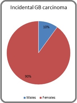
A total of 1800 Cholecystectomy cases received in the department over a period of 1 year. Ten (0.5%) cholecystectomy specimens were microscopically diagnosed as incidental gall bladder carcinoma. There were 1 (10%) male and 9 (90%) females with a Male: Female ratio of 1:9. Among total specimens obtained for histopathological examination, Chronic cholecystitis was the most frequently diagnosed accounting for 97% cases. The cases found to be malignant were further studied based on preoperative imaging findings, macroscopic findings, and pathological TNM staging. The age group affected was 31–65 years (mean – 47.7 years). Gross inspection of the majority specimens revealed thickening of gallbladder wall in 50% (5/10) cases followed by mucosal flattening with tiny papillary projections in the luminal wall in 30% (3/10) cases. 20% (2/10) did not show any macroscopic findings suggestive of malignancy. Majority of the cases of IGBC 60% (6/10) were associated with gallstones. On microscopic examination, all cases showed features of adenocarcinoma. 7 cases showed irregularly branched and dilated glands lined by columnar epithelial cells having enlarged nuclei, vesicular chromatin and prominent nucleoli and scant eosinophilic cytoplasm.
Among the remaining 3 cases, solid sheets of malignant cells (1 case), extracellular mucin pools (1 case) and arborising papillary architecture with fibrovascular core (1 case) were noted. Mitosis including atypical forms was seen. Lympho-vascular invasion was not seen. Perineural invasion were seen in 20% (2/10) cases. Tumor cells were seen infiltrating the lamina propria in 20% (2/10) cases (pT1a), muscularis propria in 60% (6/10) (pT1b), and serosa in the remaining 20% (2/10) cases (pT3) (Table 1 and 2), (Figure 1).
| Histopathological diagnosis | Number (n) | Percentage (%) |
| 1. Chronic cholecystitis | 1746 | 97 |
| 2. Gall bladder polyp | 7 | 0.38 |
| 3. Gall bladder carcinoma | 15 | 0.83 |
| 4. Goblet cell metaplasia | 21 | 1.16 |
| 5. Pyloric gland metaplasia | 7 | 0.38 |
| 6. Dysplasia | 4 | 0.22 |
| Total | 1800 |
| Subtypes with grading of GB carcinoma | Number (n=10) | Percentage (%) |
| 1. Well differentiated adenocarcinoma | 5 | 50 |
| 2. Moderately differentiated adenocarcinoma | 2 | 20 |
| 3. Poorly differentiated carcinoma | 1 | 10 |
| 4. Mucinous adenocarcinoma | 1 | 10 |
| 5. Papillary adenocarcinoma | 1 | 10 |
Figure 1. Proportion of Males and Females Having Incidental Gall Bladder Carcinoma.

Discussion
Incidental gall bladder carcinoma means carcinoma detected for the 1st time in patients undergoing cholecystectomy for benign lesions such as cholecystitis or cholelithiasis either during surgery or histopathological examination [9]. Females have a higher incidence of gall bladder carcinoma as compared to males.
The radiological evidences of gall bladder carcinoma are often non-specific showing gall bladder wall thickening which can be readily confused with cholecystitis instead of gall bladder carcinoma. Also, there are no established screening procedures for the same. Histopathological examination, therefore, is the gold standard for diagnosis of gall bladder carcinoma. After Incidental detection of GB carcinoma re-grossing and re-submission of sections from appropriate areas had to be done following cancer protocol. In our study 50% of the cases showed gall bladder wall thickening; rest of the cases had cholelithiasis and unremarkable gall bladder which were thin walled (<3mm) and anechoic.
Intraoperative frozen section diagnosis of whether a lesion is benign or malignant reduces the risk of requirement of subsequent surgeries following confirmed diagnosis by histopathology. Early-stage detection is potentially curative with surgical resection followed by adjuvant therapy. Unresectable or metastatic gall bladder cancers qualify for palliative care/ chemotherapy (Table 3).
| Author | Year | Incidence (%) |
| Shimizu T et al [10] | 2008 | 0.83 |
| Mitrovic F et al [11] | 2010 | 0.54 |
| Ghimire P et al [12] | 2011 | 1.28 |
| Panebianco A et al [13] | 2013 | 0.50 |
| Loannidis O et al [14] | 2013 | 0.20 |
| Waghmare RS et al [15] | 2014 | 2.59 |
| Martins-Fihlo Ed et al [16] | 2015 | 0.34 |
| Emmett CD et al [9] | 2015 | 0.25 |
| Duzkoylu Y et al [17] | 2016 | 0.20 |
| Ahn Y et al [18] | 2016 | 1.50 |
| Geramizadeh B et al [19] | 2017 | 0.37 |
| Our study | 2022 | 0.50 |
In conclusion, India is one country to have a high incidence of gall bladder malignancy. Within India, North Eastern region along with central Indian regions have higher risk of getting affected due to various environmental reasons such as soil and water contamination by industrial wastes which are identified as carcinogens. Due to non specific signs and symptoms gall bladder carcinoma may be detected incidentally on histopathological examination. The stage of tumor at which it is identified is of prognostic importance; early stage has better prognosis. Lastly, large multicentric studies are required to assess the risk factors of gall bladder carcinoma which will help the health care system to formulate strategies to reduce mortality and morbidity of the patients.
Acknowledgments
Statement of Transparency and Principals:
· Author declares no conflict of interest
· Study was approved by Research Ethic Committee of author affiliated Institute.
· Study’s data is available upon a reasonable request.
· All authors have contributed to implementation of this research.
References
- Risk factors for gallbladder cancer. An international collaborative case-control study Strom BL , Soloway RD , Rios-Dalenz JL , Rodriguez-Martinez HA , West SL , Kinman JL , Polansky M, Berlin JA . Cancer.1995;76(10). CrossRef
- Gallbladder cancer worldwide: geographical distribution and risk factors Randi G, Franceschi S, La Vecchia C. International Journal of Cancer.2006;118(7). CrossRef
- Global cancer statistics: GLOBOCAN estimates of incidence and mortality worldwide for 36 cancers in 185 countries; available online at https://gco.iarc.fr/ (Accessed on May 19, 2021) .
- Place of birth and risk of gallbladder cancer in India Mhatre SS , Nagrani RT , Budukh A, Chiplunkar S, Badwe R, Patil P, Laversanne M, et al . Indian Journal of Cancer.2016;53(2). CrossRef
- National Cancer registry programme. Consolidated report of population based cancer registries: 2012-14 [Internet] Available online: http://ncdirindia.org/NCRP/ALL_NCRP_REPORTS/PBCR_REPORT_2012_2014/index.htm..
- Gallbladder carcinoma. PathologyOutlines.com website. https://www.pathologyoutlines.com/topic/gallbladdercarcinoma.html. Accessed January 20th, 2023 Akki A. .
- Gallbladder cancer in India: a dismal picture Batra Y, Pal S, Dutta U, Desai P, Garg PK , Makharia G, Ahuja V, et al . Journal of Gastroenterology and Hepatology.2005;20(2). CrossRef
- Gallbladder carcinoma: radiologic-pathologic correlation Levy AD , Murakata LA , Rohrmann CA . Radiographics: A Review Publication of the Radiological Society of North America, Inc.2001;21(2). CrossRef
- Routine versus selective histological examination after cholecystectomy to exclude incidental gallbladder carcinoma Emmett CD , Barrett P, Gilliam AD , Mitchell AI . Annals of the Royal College of Surgeons of England.2015;97(7). CrossRef
- Incidental gallbladder cancer diagnosed during and after laparoscopic cholecystectomy Shimizu T, Arima Y, Yokomuro S, Yoshida H, Mamada Y, Nomura T, Taniai N, et al . Journal of Nippon Medical School = Nippon Ika Daigaku Zasshi.2006;73(3). CrossRef
- Incidental gallbladder carcinoma in regional clinical centre Mitrović F, Krdzalić G, Musanović N, Osmić H. Acta Chirurgica Iugoslavica.2010;57(2). CrossRef
- Incidence of incidental carcinoma gall bladder in cases of routine cholecystectomy Ghimire P, Yogi N, Shrestha BB . Kathmandu University medical journal (KUMJ).2011;9(34). CrossRef
- Incidental gallbladder carcinoma: our experience Panebianco A, Volpi A, Lozito C, Prestera A, Ialongo P, Palasciano N. Il Giornale Di Chirurgia.2013;34(5-6). CrossRef
- Primary gallbladder cancer discovered postoperatively after elective and emergency cholecystectomy Ioannidis O, Paraskevas G, Varnalidis I, Ntoumpara M, Tsigkriki L, Gatzos S, Malakozis SG , et al . Klinicka Onkologie: Casopis Ceske a Slovenske Onkologicke Spolecnosti.2013;26(1). CrossRef
- Incidental Gall Bladder Carcinoma in Patients Undergoing Cholecystectomy: A Report of 7 Cases Waghmare RS , Kamat RN . The Journal of the Association of Physicians of India.2014;62(9).
- Prevalence Of Incidental Gallbladder Cancer In A Tertiary-Care Hospital From Pernambuco, Brazil Martins-Filho ED , Batista TP , Kreimer F, Martins ACA , Iwanaga TC , Leão CS . Arquivos De Gastroenterologia.2015;52(3). CrossRef
- Incidental gallbladder cancers: Our clinical experience and review of the literature Düzköylü Y, Bektaş H, Kozluklu ZD . Ulusal Cerrahi Dergisi.2016;32(2). CrossRef
- Incidental gallbladder cancer after routine cholecystectomy: when should we suspect it preoperatively and what are predictors of patient survival? Ahn Y, Park CS , Hwang S, Jang HJ , Choi KM , Lee SG . Annals of Surgical Treatment and Research.2016;90(3). CrossRef
- Incidental Gall Bladder Adenocarcinoma in Cholecystectomy Specimens; A Single Center Experience and Review of the Literature Geramizadeh B, Kashkooe A. Middle East Journal of Digestive Diseases.2018;10(4). CrossRef
License

This work is licensed under a Creative Commons Attribution-NonCommercial 4.0 International License.
Copyright
© Asian Pacific Journal of Cancer Biology , 2023
Author Details