A Rare Case Report of Carcinoma Gall Bladder with Brain Metastasis in 18 Year Old Male
Download
Abstract
Introduction: Worldwide carcinoma gall bladder is 22nd most common cancer and 17th most common cause of death according to GLOBOCAN 2018 data. In 2018, the estimated incidence was 97,000 for men and 122,000 for women. Risk of carcinoma gall bladder from birth till 74 year is 0.26% for women and 0.25% for men. Here we are reporting a case of carcinoma gall bladder of 18 year old male who presented to us in opd with extensive brain metastasis and liver metastasis.
Case presentation: This case report refers to 18 year old male presented to us with chief complaint of pain in right hypochondrium for 20 days and history of headache and vomiting since 4 days. CECT whole abdomen suggestive of a 5.9*3.9*3.2 mass arising from body of gall bladder extending into segment V of liver. CEMRI BRAIN suggestive of 3.2*3 cm lesion in left frontoparietal region of brain parenchyma. USG guided FNAC from gall bladder suggestive of poorly differentiated adenocarcinoma. Patient was treated by whole brain RT on Co-60 by German helmet technique planned 30Gy/10#.
Conclusion: This case adds to our knowledge that Gall bladder cancer is extremely rare malignancy in children. These tumors are not associated with gall stones in this age group and further studies must be done to evaluate the risk factors in younger age group.
Introduction
Worldwide carcinoma gall bladder is 22nd most common cancer and 17th most common cause of death according to GLOBOCAN 2018 data [1]. Carcinoma gall bladder is a deadly cancer as it is mostly diagnosed when it has metastasized to other organs. Gall bladder constitutes 1.2% of all the cancer burden [1]. In 2018, the estimated incidence was 97,000 for men and 122,000 for women. Risk of carcinoma gall bladder from birth till 74 year is 0.26% for women and 0.25% for men [1].
Here we are reporting a case of carcinoma gall bladder of 18 year old male who presented to us in opd with extensive brain metastasis and liver metastasis.
Case Report
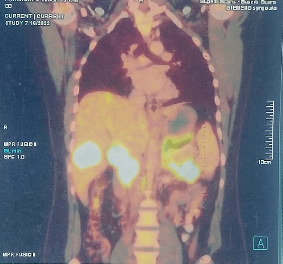
An 18 year old male presented to us with chief complaint of pain in right hypochondrium for 20 days and history of headache and vomiting since 4 days. On examination patient is well oriented to time, place and person pallor is present clinically his Eastern Cooperative Oncology Group (ECOGP) score 3. Per abdomen tenderness is present in right hypochondrium. CECT whole abdomen dated 1/7/22 suggestive of gall bladder wall 8mm in thickness. A 5.9*3.9*3.2 mass arising from body of gall bladder extending into segment V of liver.7.5*8.2*8 cm lesion in distal body and tail of pancreas. PET CT was done which further confirms 8.6*4.7*4.2 cm in gallbladder with infiltration into liver segment V (SUV max: 16.3) (Figure 1).
Figure 1. Pet CT Showing Hypermetabolic Activity with Increased SUV Uptake in Gall Bladder and Liver and Lymph Nodes .

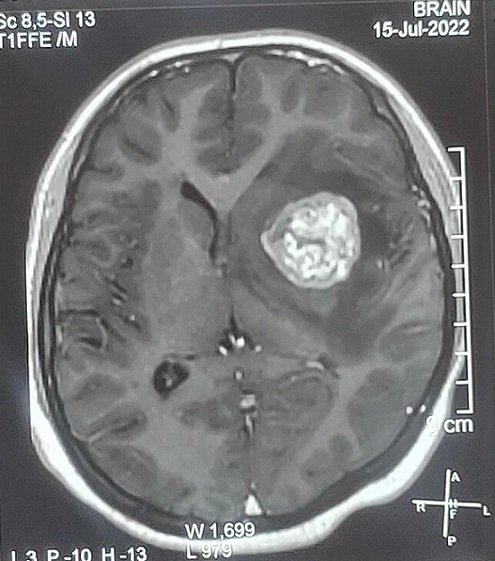
Hypermetabolic lymphatic (SUV max: 7.9), omental deposits. CEMRI BRAIN dated15/7/22 suggestive of 3.2*3 cm lesion in left frontoparietal region of brain parenchyma (Figure 2).
Figure 2. CEMRI BRAIN Showing 3.2*3 cm Lesion in Left Frontoparietal Region of Brain Parenchyma .

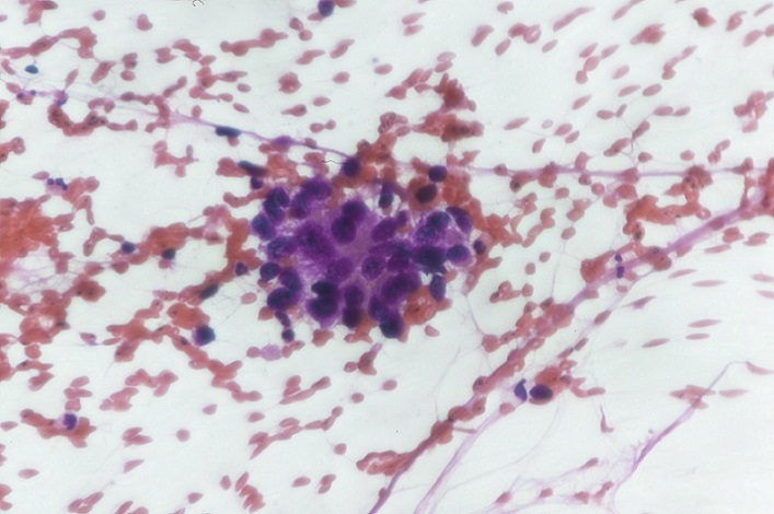
USG guided FNAC dated 9/7/22 from gall bladder suggestive of poorly differentiated adenocarcinoma (Figure 3).
Figure 3. USG Guided FNAC from Gall Bladder Suggestive of Poorly Differentiated Adenocarcinoma. (X40).

CEMRCP was done there is heterogeneously enhancing GB mass 6.4*4.4 cm with intrahepatic biliary infiltration and mild to moderate bilobar IHBRDs. Multiple liver SOL’s. No pancreaticobiliary duct abnormality was detected. Patient was treated by whole brain RT on Co-60 by German helmet technique planned 30Gy/10# was started on 17/7/22 to 27/7/22 uneventfully. He was given one cycle of gemcitabine and cisplatin subsequent cycle was deferred in view of increased serum bilirubin (17) and pt underwent Percutaneous Transhepatic Biliary Drainage (PTBD).
Discussion
Gall bladder cancer is common in sixth and seventh decade. The Surveillance, Epidemiology, and End Results (SEER) database from the US from 2015 reveals that age-adjusted incidence rates (per 100,000) in 2015 were 0.2 for those aged 20-49 years, 1.6 for those aged 50-64 years, 4.3 for those aged 65-74 years, and 8.1 for individuals aged 75 years and older [2]. Our patient is a 17year old male who presented to us in opd. As per SEER database incidence rates of carcinoma gall bladder in 20-49 years is 0.2 and rare cases have been reported in literature of gall bladder incidence in patient below 20years of age especially male patients [3,4].
Gall bladder is often diagnosed at late stage where the intent is palliative care. Gall bladder cancer is more common in women due to high estrogen and higher incidence of gall stones. A history of gallstones carries the highest risk for gallbladder cancer, with the relative risk (RR) being 4.9. In all cases of gall bladder cancer reported in children, there was no association of gall stones which has been considered as the most common risk factor for gall bladder cancer [5]. In male abnormal pancreaticobiliary junction which occur at a younger age, have been one of the cause of gall bladder cancer [6]. Our patient is 18 year old male with no history of gall stones or abnormal pancreaticobiliary duct.
Risk factors for carcinoma gall bladder are old age, obesity, female gender, genetics, occupational exposure to mutagens and chronic infection (Salmonella, Helicobacter) medications such as methyldopa, oral contraceptives pills, isoniazid, and estrogen [5]. KRAS, P16, c-erb-B2, and TP53 mutation are associated with gall bladder cancer. Gallbladder cancer in those with an anomalous pancreaticobiliary duct junction frequently presents with KRAS mutations and while in patients with cholelithiasis and chronic cholecystitis mostly present with p53 mutation.
Distant metastasis to brain occur rarely in carcinoma gall bladder and only few cases has been reported in literature [7]. Although few cases of brain metastasis has been reported in carcinoma gall bladder patient especially in younger patient but on contrary our case report is on one of this rare metastatic site. The median survival of 3 to 12 month has been reported in brain metastasis patient in carcinoma gall bladder [8].
In conclusion, Gall bladder cancer is extremely rare malignancy in children and mostly present with distant metastasis which further carries poor prognosis making the intent of treatment as palliative. These tumors are not associated with gall stones in this age group and further studies must be done to evaluate the risk factors in younger age group.
Acknowledgments
Statement of Transparency and Principals:
· Author declares no conflict of interest
· Study was approved by Research Ethic Committee of author affiliated Institute.
· Study’s data is available upon a reasonable request.
· All authors have contributed to implementation of this research.
References
- Global cancer statistics 2018: GLOBOCAN estimates of incidence and mortality worldwide for 36 cancers in 185 countries Bray F, Ferlay J, Soerjomataram I, Siegel RL , Torre LA , Jemal A. CA: a cancer journal for clinicians.2018;68(6). CrossRef
- Epidemiology of colorectal cancer: incidence, mortality, survival, and risk factors Prashanth Rawla , Tagore Sunkara , Adam Barsouk . Gastroenterology Rev .2019;14(2):89-103. CrossRef
- Cancer of the gallbladder in an 11-yr-old Navajo girl Rudolph R, Cohen JJ . Journal of Pediatric Surgery.1972;7(1). CrossRef
- Cancer of the gallbladder in a 9-year-old girl Iwai N, Goto Y, Taniguchi H, Tokiwa K, Tsuto T, Yanagihara J, Takahashi T, Majima S, Kano M. Zeitschrift Fur Kinderchirurgie: Organ Der Deutschen, Der Schweizerischen Und Der Osterreichischen Gesellschaft Fur Kinderchirurgie = Surgery in Infancy and Childhood.1985;40(2). CrossRef
- Epidemiologic aspects of gallbladder cancer: a case-control study of the SEARCH Program of the International Agency for Research on Cancer Zatonski WA , Lowenfels AB , Boyle P, Maisonneuve P, Bueno de Mesquita HB , Ghadirian P, Jain M, et al . Journal of the National Cancer Institute.1997;89(15). CrossRef
- Japanese clinical practice guidelines for pancreaticobiliary maljunction Kamisawa T, Ando H, Suyama M, Shimada M, Morine Y, Shimada H. Journal of Gastroenterology.2012;47(7). CrossRef
- Brain metastasis as an initial manifestation of of gallbladder carcinoma Joel R. Tayo , Huda Al-Abdulkarim , Molham Al-Rayess . Neurosciences.2005;10(3):235-237.
- The management of cerebral metastasis Lokich JJ . JAMA.1975;234(7).
License

This work is licensed under a Creative Commons Attribution-NonCommercial 4.0 International License.
Copyright
© Asian Pacific Journal of Cancer Biology , 2023
Author Details