Rectal Polyp Prolapse: A Case Report
Download
Abstract
Colorectal adenomas are polyps that develop from the mucosa and exhibit neoplastic characteristics. Adenomas’ increasing dysplasia and malignant potential are connected to their size, villous content, and patient’s age. An anorectal emergency is definitely a possibility when there are large villous polyps in the rectum. They could be involved in rectal bleeding, blockage, prolapse, or imprisonment. We describe a 53-year-old female who was treated successfully for giant tubulovillous rectal adenoma that was prolapsed through anal opening. The patient’s clinical symptoms and signs were mistaken for prolapsed hemorrhoids.
Introduction
Histologically, colorectal polyps can be classed as neoplastic (which can be benign or malignant), adenomatous (including serrated adenomatous) or non-neoplastic (including hyperplastic, mucosal, inflammatory and hamartomatous) polyps. By the ages of 50 and 70, the general population is affected by adenomatous polyps in around 33% and 50%, respectively, of the population. A single adenoma is present in 60% of cases, whereas numerous lesions are present in 40% of cases. The majority of lesions are less than 1 cm in size. Lesions will be found 60% of the time distal to the splenic flexure [1]. In an emergency situation, prolapsing anorectal polyps can mimic benign anorectal diseases like prolapsed hemorrhoids and provide treatment challenges.
Case Report
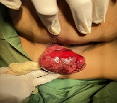
A 53-year-old female patient was admitted to our emergency department with a mass protruding from the anal canal. She had a prolapsing rectal mass approximately for two years, although she always refused further colonoscopic evaluation or surgical treatment since the mass was relocated spontaneously. On admission, she did not refer to abdominal pain or diarrhea. She mentioned chronic constipation and rarely the urgency of defecation. Physical examination did not reveal abdominal pain or signs of intestinal obstruction. There were no symptoms of intestinal obstruction or abdominal discomfort upon physical examination. A prolapsed mass with a diameter of 7 cm that was discovered during rectal examination while the patient was in the lithotomy posture (Figure 1).
Figure 1. A Prolapsed Mass with a Diameter of 7 cm.

The bulk had a rotting surface, erosion, and a bad odor. The other biochemical readings were normal, and the hemoglobin level was 7.6 g/dL.Under general anesthesia, the polyp that was protruding from the anal canal was removed via transanal excision. By transanally excising the bulk and underlying muscle layer in one piece, clean surgical margins were achieved. There were no difficulties after the operation. The pathologic evaluation of the tumor revealed that it was a tubulovillous adenoma with intramucosal carcinoma. No lymphovascular invasion was seen, and the tumor was ow-grade and well-to-moderately differentiated. No further treatment was recommended.
Discussion
There are polyps in each part of the colon. Adenomatous polyps can have one of three basic histologic subtypes: tubular, villous, or tubulovillous. The World Health Organization defines tubular adenomas as having less than 25% villous component, 25-75% tubulovillous component, and greater than 75% villous component [2]. The most frequent types of adenomas are tubular, tubulovillous, and villous. There are equal numbers of tubular adenomas throughout the colon. The rectum is a preferred location for villous adenomas. They could be asymptomatic or connected to bleeding, blockage, electrolyte imbalance, mucous excretion, or diarrhea [2,3]. Villous adenomas have a chance of 35–40%, tubulovillous adenomas of 20–25%, and tubular adenomas of 5% [4]. The management of polyps should be based on the size and shape of the polyps, which are two critical characteristics that may indicate the presence of underlying malignancy. They might be sessile (typically tubulovillous or villous), pedunculated (typically tubular or tubulovillous), or non-polypoid (flat or depressed). Only 1.3% of small adenomas (less than 1 cm) are malignant, whereas 46% of adenomas larger than 2 cm are. On complete removal of the polyp, malignant cells are discovered in 5.7%, 18%, and 34.5% of adenomatous polyps with mild, moderate, and severe dysplasia, respectively [1]. According to the United States National Polyp Study, an advanced adenoma is one that is less than one centimeter in size or contains invasive malignancy or high-grade dysplasia. It has repeatedly been demonstrated that a negative resection margin is linked to a lower likelihood of unfavorable outcomes (recurrence, residual carcinoma, lymph node metastases, and shorter survival). The pathologic analysis revealed that the polyp in our patient was a tubulovillous polyp with intramucosal carcinoma in the polyp head and was excised with a negative resection margin. The stem of the polyp was not invaded. Polyps that prolapse through the anus are associated by a number of different ways. Since there is less fat in the ischiorectal fossa during the earliest years of life, less pressure is applied to this essential part of the perineum, it appears that this condition is more common in children [5-7]. Adults are most susceptible to prolapse when their anal sphincter is damaged or dysfunctional or when they have illnesses like chronic constipation that raise intra-abdominal pressure [8]. In our case, the patient experienced constipation but no obvious changes in the integrity of the anal sphincter that would lead to prolapse. Treatment options for prolapsed polyps include conservative treatment, endoscopic resection, and even ultralow anterior excision, albeit there is no universally accepted method [9]. Nearly all pedunculated polyps can be removed safely and effectively using colonoscopic polypectomy. Finally, screening and surveillance programs are advised since people with anorectal adenomatous polyps have a known higher risk of developing cancer. If the entire polyp was removed without partial removal, patients with 3-10 adenomas, any adenoma less than 1 cm, any adenoma with villous features, or high-grade dysplasia should have their next colonoscopy in 3 years [10,11].
Acknowledgments
Statement of Transparency and Principals:
· Author declares no conflict of interest
References
- Cancer of the Colon, Rectum, and Anus. In: Clarke CN, You YN, Feig BW, eds. The MD Anderson Surgical Oncology Handbook (6th ed.). Philadelphia; Wolters Kluwer Feig BW , Ching CD . 2019;:491-457.
- Carcinoma of the colon. Charles C McKittrick LS , Wheelock FC Jr . Thomas; Springfield, IL.1954;:61-63.
- Combined restorative proctocolectomy and pancreaticoduodenectomy for familial adenomatous polyposis Jatzko G, Siebert F, Wolf B, Karner-Hanusch J, Kleinert R, Denk H. Zeitschrift Fur Gastroenterologie.1999;37(11).
- Colorectal Cancer: Epidemiology, Risk Factors, and Health Services Amersi F, Agustin M, Ko CY . Clinics in Colon and Rectal Surgery.2005;18(3). CrossRef
- Peutz-Jeghers syndrome in a child. Prolapse of a large colonic polyp through the anus Mönig SP , Selzner M, Schmitz-Rixen T. Journal of Clinical Gastroenterology.1997;25(4). CrossRef
- An unusual hamartomatous malformation of the rectosigmoid presenting as an irreducible rectal prolapse and necessitating rectosigmoid resection in a 14-week-old infant Lamesch AJ . Diseases of the Colon and Rectum.1983;26(7). CrossRef
- Peutz-Jeghers syndrome: its natural course and management Utsunomiya J, Gocho H, Miyanaga T, Hamaguchi E, Kashimure A. The Johns Hopkins Medical Journal.1975;136(2).
- Rectal prolapse Melton GB , Kwaan MR . The Surgical Clinics of North America.2013;93(1). CrossRef
- Ultralow anterior resection for prolapsed giant solitary rectal polyp of Peutz-Jeghers type Garces M, García-Granero E, Faiz O, Alcacer J, Lledó S. The American Surgeon.2011;77(4).
- Recommendations for post-polypectomy surveillance in community practice Ransohoff DF , Yankaskas B, Gizlice Z, Gangarosa L. Digestive Diseases and Sciences.2011;56(9). CrossRef
- Efficacy and safety of endoscopic resection of large colorectal polyps: a systematic review and meta-analysis Hassan C, Repici A, Sharma P, Correale L, Zullo A, Bretthauer M, Senore C, et al . Gut.2016;65(5). CrossRef
License

This work is licensed under a Creative Commons Attribution-NonCommercial 4.0 International License.
Copyright
© Asian Pacific Journal of Cancer Biology , 2023
Author Details