Twenty-Seven Cases of Marjolins Ulcer; An Institutional Experience on Diagnosis, Treatment and Outcomes
Download
Abstract
Purpose: Marjolins ulcer is a malignant transformation that arises from chronic ulcers or previously traumatized scar that occur usually after burns. To study the clinicopathological characteristics and treatment outcomes of Marjolins ulcer at our institute.
Materials and methods: Retrospective analysis of all Marjolins ulcer patients presented to our department from 2018 to 2021 was done. A total of 27 patients of all age groups were included in the study. All the information regarding the diagnosis, treatment and outcome details were collected and analysed.
Results: Most of the patients were in the 5th decade of life with an overall male preponderance. The most common cause for Marjolin ulcer was Burns Scar. The mean latency period for the development of Marjolins ulcer was 11 years. Squamous cell carcinoma was the most common histological subtype. 18.5% patients received Adjuvant radiotherapy. At the median follow up of 14 months, one patient presented with locoregional relapse.
Conclusion: Chronic non-ulcers that do not respond to treatment should be carefully examined by multidisciplinary team for malignant transformation. Surgery is the mainstay of treatment and Adjuvant Radiotherapy should be considered in high-risk cases to reduce locoregional recurrence. Tumour size and nodal involvement are the main predictors of locoregional relapse.
Introduction
Marjolin’s ulcer ,a cutaneous malignancy was first described by a a French surgeon Jean Nicholas Marjolin, and he described the ulcer formation over the burns scar, although they were not recognized as malignant at that time [1]. The term Marjolin’s ulcer was defined by DaCosta, for the carcinomas arising from the burns scar [2]. Marjolin’s ulcer is considered as a highly aggressive disease that develops from chronic wounds and skin scars and almost 65% of these ulcers have been diagnosed on underlying burn scars [3]. It can also develop on discoid lupus erythematosus lesions, ulceration and chronic osteomyelitis, amputation stumps, regions of chronic fistulas, chronic wounds etc [4-7]. It can occur at any age group but it is less common in children [8]. Marjolin’s ulcer are predominantly seen in males [6,7]. Squamous cell carcinoma is the most common histologic variant other variants are basal cell carcinoma (BCC), angiosarcoma, fibrosarcoma, liposarcoma osteosarcoma etc [9].
The mechanism of malignant transformation is very well understood. A lot of theories have been mentioned in the literature [10]. Few theories states that the mutation by the inflammation in the injured tissue causes carcinogenesis [11]. Others describe that foreign body reaction at the damaged tissue leading to malignant transformation [12]. Few other studies also states that repeated damage to the ulcer and long-standing chronic irritation, which leads to continuous mitotic activity, to reduce the defect which ultimately leading to Carcinogenesis [13]. Patients with immune deficiency which are inherited are at increased risk for Carcinoma formation [14]. Marjolin’s ulcers are divided into two type acute and chronic. In acute, malignant transformation happened within one year of the injury [15] and the chronic happens occur over years of long latency time. Marjolins ulcer is confirmed by histological examination of the tissue from the damaged site. The present study is a retrospective analysis of Marjolin’s ulcer ,diagnosis, treatment and outcomes in a tertiary care hospital in India.
Materials and Methods
It was a hospital based retrospective study. All the patients presented to our institute from 2018 to 2021 with diagnosis of Marjolins ulcer were analysed. All the information related to clinical features, history, diagnosis, treatment and follow-up details were collected. Histopathological examination was considered as the gold standard for diagnosis.
All the patients were treated by surgery as the main modality of treatment. Adjuvant Radiation was given in the patients who were meeting high risk criteria. After the completion of the treatment all the patients were followed up every 3 months with clinical and radiological examination. All the details were documented and analyzed. Statistical analysis was done by using the software SPSS 22.0 and R environment version.3.2.2 and Microsoft word and Excel have been used to create tables etc. Descriptive analysis and inferential analysis have been done in the study.
Results
Total Twenty-Seven cases of Marjolin’s ulcers were identified. They were stratified according to the age, gender and the anatomic location. The age group of the patients studied ranged from 35 to 70 years. The median age of the study population was 52 years. Males were more commonly affected. Lower extremity was involved in 59% (16) of the patients followed by Upper Extremity (p 0.203). The most common etiologic factor was the burns scar followed by trauma which was statistically significant with a p value of 0.065. The time between the etiological factor and occurrence of Marjolin ulcer was 7 to 15 years (Mean 11years).Patient and tumour characteristics are explained in Table 1.
| Age | Mean 52 Years | n | Percentage (%) | P value |
| Male | 17 | 62 | ||
| Gender | Female | 10 | 38 | P=0.203 |
| Lower Extremity | 16 | 59 | ||
| Site | Upper Extremity | 5 | 18 | |
| Head and Neck | 5 | 18 | P=0.065 | |
| Trunk | 1 | 4 | ||
| Etiology | Burns | 17 | 62 | P=0.012 |
| Trauma | 9 | 33.00 | ||
| DLE | 1 | 5.00 | ||
| Histology | Squamous cell Carcinoma | 23 | 85.00 | |
| Basal Cell Carcinoma (BCC) | 3 | 10.00 | P-0.045 | |
| Dermatofibrosarcoma | 1 | 5.00 |
All the patients underwent imaging of the locoregional site to rule out locoregional nodal spread. Surgery was the main modality of treatment. Wide Local Excision with 2 cm margin was conducted as the standard of management followed by reconstructions with Split Skin Graft (SSG) or locoregional flaps.
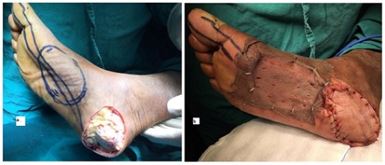
Out of the 16 lower extremity patients inguinal nodal dissection was done in 6 patients due to involvement of lymph nodes either clinically or radiologically. Four patients were presented with extensive bony involvement in which amputation followed by reconstructions with split skin graft (SSG) was conducted (Figure 1).
Figure 1. Pre- and Post-Operative Images of the Marjolins Ulcer of the Foot.

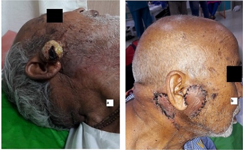
In upper extremity tumours, one patient presented with axillary nodal metastasis on presentation for which Axillary Nodal dissection was done. All the patients Marjolins ulcer in head and neck region underwent WLE with adequate margins (Figure 2).
Figure 2. Pre and Post Operative Image of the Marjolins Ulcer of the Face.

One patient developed Marjolins ulcer due to Discoid Lupus Erythematosus and he underwent wide local excision with parascapular flap reconstruction.
On Histopathological examination, the size of the tumour ranged from 2x3 cm to 10x10 cm. Squamous cell Carcinoma (SCC) (23) in most common histology followed by, Basal Cell Carcinoma (BCC) (3) and Dermatofibrosarcoma (1). Five patients were found to have positive lymph nodes post operatively.
Adjuvant Radiotherapy was given in the patients with high-risk criteria positive lymph nodes and larger tumour size. Five patients were received adjuvant Radiotherapy. All the patients were followed every 3 months with clinical and radiological examination of primary site and nodal areas. At the median follow-up of 14 months, all the patients showed complete response except one patient. This patient with SCC of the foot came back with inguinal lymph nodal recurrence after 1 year of surgery. The Progression Free Survival at 3 years was found to be 96. 3% with a significant p value of 0.035.
Discussion
Non-healing ulcers which were developed on the chronic scars are very dangerous because of their malignant transformation potential. Hence, these ulcers should be examined carefully to exclude the presence of malignancy. Clinical profile of both benign and malignant ulcers is same, although some variations are noted [16,17]. Hence, any ulcer which is not healing on burns or traumatic scar should be treated like a cancerous one unless it is proved by histological examination [18]. This retrospective analysis was done to study the clinicopathological profile and treatment outcomes of Marjolins ulcers presented at our institute.
Marjolins ulcers may occur at any age with no strong race predisposition [19]. It is most commonly seen in men and this ratio in our study, was 1.9:1. Men are at major risk for developing Marjolins ulcer may be due to the genetic mutations and more physical activities by the males. The mean age of our study cases was 52 years with an age range from 35 to 70 years. Which is identical to other studies [20,17]. Most frequently observed site was lower extremity in our study. Marjolin’s ulcer may occur on any site , but they are commonly observed at lower limbs followed by upper limbs, head and neck and trunk [21]. Our objective was to analyse the clinicopathological profile and treatment outcomes in the biopsy proven Marjolin’s ulcer. In our study, squamous cell carcinoma (SCC) was the most common histological subtype. These findings are in an agreement with literature [22] We recorded the cutaneous ulcers within the scars in the majority of the cases 23 out of 27, fungating ulcers in 4 patients, clinical or radiologically significant lymphadenopathy was found in six patients. The extensive bony involvement was found in four cases. The mean dimension of the ulcers in our study was 6-8 cm (ranging from 2 to 12 cm) According to literature, ulcers with more than 6 cm are more likely to undergo malignant transformation [23].
Marjolin’s ulcer rarely metastasize. It is generally considered that prophylactic nodal dissection has no influence on recurrence and should not be encouraged [24]. However, few studies reported that, of lymph nodes spread in the lower limbs is relatively more and for the patients without lymph node spread, prophylactic nodal dissection should be considered [25]. Few studies also say that Lymph node metastasis in Marjolins ulcer is 22% [26]. Lymph nodal involvement in our study was 22%.
Surgery is the standard treatment method for Marjolin’s ulcer. Post excision of the tumour, repair and reconstruction using skin grafts to be done to improve the quality of life. Skin grafting should be considered as much as possible. skin flap repair should be considered if bones are exposed. Local skin flap repair should be done, if possible, otherwise skin flap graft repair is the considered as an alternative option.
The histological subtype SCC has a worst prognosis compared to other types, hence aggressive treatment in this subtype is encouraged-excision and radiotherapy are recommended for managing the recurrence. A study done by Ozek and Cankayal found that the radiation should be given in patients positive lymph nodes after nodal dissection, tumours with more than 10 cm. In our study adjuvant radiation was given in the patients with positive nodes. Overall, literature reviews support the use of adjuvant radiation in poor surgical candidates, positive nodes and large sized tumours [27].
Marjolin’s ulcer has a very short recurrence time [28,29]. However, recurrence rates in our study did not show any statistical significance. The main reason for this is the smaller number of patient population and the shorter duration of follow-up.
In conclusion, Chronic non-ulcers that do not respond to treatment should be carefully examined by multidisciplinary team for malignant transformation. This retrospective review at our institute showed that Marjolin’s ulcer predominantly seen in males and the most common etiological factor was burns scar. Surgery followed by Adjuvant Radiation should be considered in high-risk patients.
Acknowledgments
Statement of Transparency and Principals:
· Author declares no conflict of interest
· Study was approved by Research Ethic Committee of author affiliated Institute.
· Study’s data is available upon a reasonable request.
· All authors have contributed to implementation of this research.
References
- Marjolin's ulcer: a preventable complication of burns? Copcu E. Plastic and Reconstructive Surgery.2009;124(1). CrossRef
- Marjolin's ulcer arising in a burn scar Dupree MT , Boyer JD , Cobb MW . Cutis.1998;62(1).
- A Comprehensive Review on Marjolin's Ulcers: Diagnosis and Treatment Pekarek B, Buck S, Osher L. The Journal of the American College of Certified Wound Specialists.2011;3(3). CrossRef
- Squamous cell carcinoma complicating discoid lupus erythematosus in Chinese patients: review of the literature, 1964-2010 Tao J, Zhang X, Guo N, Chen S, Huang C, Zheng L, Li Y, et al . Journal of the American Academy of Dermatology.2012;66(4). CrossRef
- Marjolin's ulcer associated with ulceration and chronic osteomyelitis Tavares E, Martinho G, Dores JA , Vera-Cruz F, Ferreira L. Anais Brasileiros De Dermatologia.2011;86(2). CrossRef
- Marjolin's ulcer in an amputation stump Bloemsma GC , Lapid O. Journal of Burn Care & Research: Official Publication of the American Burn Association.2008;29(6). CrossRef
- Marjolin's ulcer: Sequelae of mismanaged chronic cutaneous ulcers Asuquo ME , Ikpeme IA , Ebughe G, Bassey EE . Advances in Skin & Wound Care.2010;23(9). CrossRef
- Marjolin's ulcer: clinical and pathologic features of 83 cases and review of literature Sadegh Fazeli M, Lebaschi AH , Hajirostam M, Keramati MR . Medical Journal of the Islamic Republic of Iran.2013;27(4).
- Marjolin ulcer: a case report Ortiz BDM , Riveros R, Gabriela MB , R M, Masi MR , Knopfelmacher O, Lezcano LB . Nasza Dermatologia Online.2014;5(1). CrossRef
- Marjolin’s ulcer of the thigh after burn injury- case report and review of literature Wronski K. New Medicine.2014;18:69-75.
- Marjolin's ulcers: theories, prognostic factors and their peculiarities in spina bifida patients Nthumba PM . World Journal of Surgical Oncology.2010;8. CrossRef
- The development of cancer in burn scars: an analysis and report of thirty-four cases Treves N. Surg Gynecol Obstet.1930;51:749-82.
- Trauma and skin cancer: Implantation of epidermal elements and possible cause Neuman Z, Ben-Hur N, Shulman J. Plastic and reconstructive surgery.1963;32:649-656.
- Clinical study of Marjolin's ulcer Hahn SB , Kim DJ , Jeon CH . Yonsei Medical Journal.1990;31(3). CrossRef
- An overview of heel Marjolin's ulcers in the Orthopedic Department of Urmia University of Medical Sciences Shahla A. Archives of Iranian Medicine.2009;12(4).
- Early Marjolin's ulcer developing in a penile human bite scar of an adult patient presenting at Bugando Medical Centre, Tanzania: A case report Chalya HL , Mabula JB , Gilyoma JM , Rambau P, Masalu N, Simbila S. Tanzania Journal of Health Research.2012;14(4).
- MALIGNANCY IN SCARS, CHRONIC ULCERS, AND SINUSES Cruickshank AH , Mcconnell EM , Miller DG . Journal of Clinical Pathology.1963;16(6). CrossRef
- Carcinoma Within A Chronic Burn Scar (Marjolin's Ulcer). Report Of A Case Geller SA , Kurtz LH , Spellman MW . The Medical Annals of the District of Columbia.1964;33.
- Marjolin's ulcers at a university teaching hospital in Northwestern Tanzania: a retrospective review of 56 cases Chalya PL , Mabula JB , Rambau P, Mchembe MD , Kahima KJ , Chandika AB , Giiti G, et al . World Journal of Surgical Oncology.2012;10. CrossRef
- Is surgery an effective and adequate treatment in advanced Marjolin's ulcer? Aydoğdu E, Yildirim S, Aköz T. Burns: Journal of the International Society for Burn Injuries.2005;31(4). CrossRef
- (PDF) Marjolin’s Ulcer: Mismanaged Chronic Cutaneous Ulcers Asuquo ME , Nwagbara VI , Omotoso A , et al . J Clin Exp Dermatol Res S.2013;6:228-239.
- [Marjolin's ulcer in chronic osteomyelitis: seven cases and a review of the literature] Bauer T, David T, Rimareix F, Lortat-Jacob A. Revue De Chirurgie Orthopedique Et Reparatrice De L'appareil Moteur.2007;93(1). CrossRef
- Squamous cell carcinoma arising in osteomyelitis and chronic wounds. Treatment with Mohs micrographic surgery vs amputation Kirsner RS , Spencer J, Falanga V, Garland LE , Kerdel FA . Dermatologic Surgery: Official Publication for American Society for Dermatologic Surgery [et Al.].1996;22(12). CrossRef
- Burn scar carcinoma with longer lag period arising in previously grafted area Türegün M, Nişanci M, Güler M. Burns: Journal of the International Society for Burn Injuries.1997;23(6). CrossRef
- Marjolin's ulcer Battaglia M, Treadwell T. The Alabama Nurse.2008;35(4).
- Marjolin's ulcers arising in burn scars Ozek C, Cankayali R, Bilkay U, Guner U, Gundogan H, Songur E, Akin Y, Cagdas A. The Journal of Burn Care & Rehabilitation.2001;22(6). CrossRef
- Squamous carcinoma in scars: clinicopathological correlations Visuthikosol V, Boonpucknavig V, Nitiyanant P. Annals of Plastic Surgery.1986;16(1). CrossRef
- Marjolin's ulcer--a diagnostic dilemma Agale SV , Kulkarni DR , Valand AG , Zode RR , Grover S. The Journal of the Association of Physicians of India.2009;57.
- [Cancers arising from burn scars: 62 cases] Jellouli-Elloumi A, Kochbati L, Dhraief S, Ben Romdhane K, Maalej M. Annales De Dermatologie Et De Venereologie.2003;130(4).
License

This work is licensed under a Creative Commons Attribution-NonCommercial 4.0 International License.
Copyright
© Asian Pacific Journal of Cancer Biology , 2024
Author Details