Eyelid Tumours: An Institutional Experience on Clinicopathological Profile and Management
Download
Abstract
Background: Primary cancer of the eyelid is an uncom¬mon malignancy with a metastatic potential. The objective of this study to assess the clinicopathological profile, management management strategies and build awareness about the more aggressive eyelid malignancies to reduce morbidity and mortality.
Methods: Retrospective analysis of all the eyelid tumours presented to our institute from 2015 to 2021 was done. A total of 10 patients with histopathologically proven malignant eyelid tumours of all age groups were included in the study. All information regarding clinical details, treatment and outcomes were retrospectively collected and analysed.
Results: The most common malignant eyelid tumour was basal cell carcinoma (n=5) followed by squamous cell carcinoma (n=3), Malignant Melanoma (n=1) and sebaceous gland carcinoma (n=1). Mean age of all patients with malignant eyelid tumour at the time of diagnosis was 61 years. Females were more frequently affected than males. The proportion of involvement of lower eyelid was significantly higher than of upper eyelid in basal cell carcinoma (P = 0.045). All patients were managed by surgical excision with tumour-free margins verified on histopathology followed by eyelid reconstruction and adjuvant radiotherapy was given in patients meeting high risk criteria.
Conclusions: Basal cell carcinoma was the most common eyelid malignancy observed and is more frequent in women than in men. Surgery is the mainstay of treatment and adjuvant radiation in high-risk patients provides excellent locoregional control.
Introduction
Eyelid and peri-ocular skin lesions are very common in patients. These lesions are numerous due to the unique anatomical features of the eyelid as all the skin structures and its appendages, muscle, modified glands, and conjunctival mucous membrane are represented in the eyelid [1]. Eyelid malignancies are rare, representing 3% of all skin cancers in the head and neck region [2]. Their importance lies in their special site with the ability to penetrate all layers of the eyelid. These destructive lesions, by involving the lid margin or lachrymal system, can produce severe functional disability and in addition can be very disfiguring. Eyelid lesions are often misdiagnosed and lead to recurrences of the disease. Hence, the accuracy of diagnosis and definitive treatment depends on histopathological diagnosis [3]. The most common primary eyelid malignancy is BCC which is rarely metastatic. Other carcinomas such as squamous cell carcinoma (SCC) is the second most common eyelid tumour, Sebaceous gland carcinoma, malignant melanoma, Markel cell carcinoma accounting for most of the remainder of eyelid malignancies, they are associated with more spreading in nature to the surrounding structures and a more pronounced metastatic potential [4-7].
Management of eyelid malignancy consists of Mohs micrographic surgery or wide excision with negative microscopic margin clearance followed by eyelid reconstruction [8]. Intraoperative microscopic evaluation of surgical margins by frozen section results in excellent rates of local control for basal cell carcinomas and squamous cell carcinomas of the eyelid and periocular structures [9-11]. Adverse prognostic features include involvement of the upper eyelid, a tumour size of 10 mm or more, and a duration of symptoms of over six months. Adjuvant radiotherapy is advised for the patients with eyelid with residual disease, positive or close margins, lymph node involvement, lymphovascular invasion, perineural invasion or deep muscle invasion to increase the likelihood of locoregional control [12-14]. Patients with locally advanced BCC who are not amenable to definitive surgery, newer targeting therapies, or systemic treatments may be the alternative options to preserve the globe [15, 16].
In the current study, our goals were to retrospectively evaluate the clinicopathological profile, management and outcomes in patients with eyelid tumours presented to our institute.
Materials and Methods
It was a hospital based retrospective study. All the patients with eyelid malignancies presented to our institute from 2015 to 2021 were included in the study. Histopathological examination was considered as the gold standard for the diagnosis. All the information related to clinical details, treatment and follow-up details were collected and analysed. All the patients were treated by surgery as the main modality of treatment. Adjuvant Radiation was given in the patients who were meeting high risk criteria. After the completion of the treatment all the patients were followed up every 3 months with clinical and radiological examination. Statistical analysis was done by using the software SPSS 22.0 and R environment version.3.2.2 and Microsoft word and Excel have been used to create tables etc. Descriptive analysis and inferential analysis have been done in the study.
Results
Total Ten cases of Eyelid Tumours were identified. They were stratified according to the age, gender and the anatomic location. The age group of the patients studied ranged from 35 to 70 years. The median age of the study population was 61 years. Females were more commonly affected. Lower eyelid was involved in 70% (n=7) of the patients (p 0.001). Patient and Tumour Characteristics are given in Table 1.
| Age | Mean 61 Years | n | Percentage | P value |
| Male | 4 | 40 | ||
| Gender | Female | 6 | 60 | P=0.045 |
| Upper Eyelid | 3 | 30 | ||
| Site | Lower Eyelid | 7 | 70 | P=0.001 |
| Histology | Basal Cell Carcinoma (BCC) | 5 | 50 | |
| Squamous cell Carcinoma (SCC) | 3 | 30 | P-0.045 | |
| Malignant Melanoma | 1 | 10 | ||
| Sebaceous Gland Carcinoma | 1 | 10 |
Histopathological examination was considered as the gold standard for diagnosis. All the patients underwent Surgical Excision as the main modality of treatment.
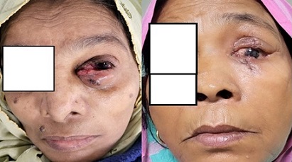
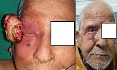
Patients with regional nodal metastasis at the time of diagnosis of the primary eyelid or conjunctival tumour underwent completion neck dissection. Preoperative and post operative images of the patients are given in Figure 1 and Figure 2.
Figure 1.A 64-year-old Female Presented with BCC of Lower Eyelid (Pre-op), for which She Underwent Surgical Excision (Post Op).

Figure 2. A 76-year-old Male Presented with SCC of Upper Eyelid (Pre-op) and he Underwent Surgical Excision (Post Op).

Five patients had postoperative adjuvant EBRT because of aggressive histologic subtypes recurrent tumour after previous failed surgical excision (1 patient), microscopic perineural invasion in the surgical specimen (1 patients), advanced- stage disease (i.e., presence of regional nodal metastasis at time of diagnosis of primary tumour) (1 patient), and microscopically positive surgical margins (2 patients) associated with a high risk of recurrence. Summary of the cases are described in Table 2.
| Case | Demographics | Eyelid | Diagnosis | Treatment | Follow-up |
| 1 | 55/F | Lower | BCC | Surgical Excision+ RT | NED |
| 2 | 75/F | Upper | SCC | Surgical Excision | NED |
| 3 | 76/M | Lower | SCC | Surgical Excision+ RT | NED |
| 4 | 68/F | Upper | BCC | Surgical Excision+ RT | NED |
| 5 | 35/M | Lower | BCC | Surgical Excision+ RT | NED |
| 6 | 64/F | Lower | BCC | Surgical Excision | Recurrence at 1 year |
| 7 | 65/M | Upper | Sb GC | Surgical Excision | NED |
| 8 | 62/F | Lower | SCC | Surgical Excision+ RT | NED |
| 9 | 55/M | Lower | MM | Surgical Excision | NED |
| 10 | 68/F | Lower | BCC | Surgical Excision | NED |
BCC; Basal Cell Carcinoma, SCC; Squamous cell Carcinoma, Sb GC; Sebaceous Gland Carcinoma. NED; No Evidence of Disease, RT; Radiotherapy.
Different types of radiation were used: 3 patients received electrons; 1 patient received photons; and 1 patient received a combination of electrons and photons. Intensity-modulated EBRT was used in 2 patients. The total radiation dose ranged from 60-66 Gy (median, 60 Gy) divided in fractions of 2 Gy per session 5 sessions per week. Additionally, skin bolus in the form of tissue equivalent material was used to ensure that the dose to the postoperative bed was optimized, and the thickness of the bolus material varied depending on the beam energy. Four patients had radiation-induced ocular side effects. All experienced some degree of dry eye syndrome, 3 had keratinization of the conjunctiva. The majority of ocular side effects of EBRT were managed conservatively with frequent lubrication, eyelid hygiene, topical medications. After the completion of treatment, patients were followed up at every 3 months intervals. All patients were followed up every 3 months with clinical and radiological examination of primary site and nodal areas. At the median follow up 20 months one patient developed a local recurrence. Initially, this patient had a aggressive histological subtype, metatypical histology. She was presented with extensive disease on recurrence and underwent orbital exenteration with adjuvant radiation. Relapse Free Survival (RFS) in our study was found to be 90% at the end of 3 years (p-0.06).
Discussion
Eyelid lesions can be benign or malignant, and hence should be examined for malignant changes like ulceration, crusting, fine telangiectatic vessels, loss of lashes, irregular pigmentation, loss of eyelid architecture, induration of edges, fixation to underlying tissue and enlarged regional lymph nodes. In this study, the mean age was 61 years with a minimum age of 35 years and a maximum age of 75 years. Malignant eyelid tumours are rare in children and young adults but occur more commonly in the sixth, seventh, and eighth decades of life [17-24]. The incidence of BCC is higher in people over 60 years of age [25].
The diagnosis of eyelid tumour was confirmed by histopathological analysis with a correlation of clinical findings. Lymph node metastasis was noted in only in one patient of BCC and Malignant melanoma. Lymph node metastasis is more common with malignant melanoma, Merkel cell carcinoma, and lymphoma [18, 26, 27]. BCC (85–95%) is the most common eyelid carcinoma, whereas others eyelid malignancies accounts 5–15% [5, 28-32, 25, 33]. Squamous cell carcinoma (SCC) accounts for 3.4 to 12.6% of eyelid cancers [34] and was the second histological type after BCC in our study [34]. The most common are the lower eyelid and the inner canthus [35].
Management of eyelid malignancies requires different considerations from other cutaneous malignancies due to their location in the periocular region and the functional impact of complete surgical resection on ocular protection and visual function. Many factors may influence the therapeutic decision such as the age of the patient, its general status, comorbidities, tumour location and stage, histological type and prior treatments. Surgical excision with the minimal recommended surgical margins of 3-4 mm was the main modality of treatment in our study. The standard modality for biopsy technique is frozen section control excision biopsy or Moh’s micrographic surgery control [17, 36]. More important margins of 5-10 mm are needed in recurrent BCC, in nodular subtype if the tumour size exceeds 1cm, infiltrative subtype [35]. Eyelid reconstruction is performed depending on eyelid defects following surgical excision. by direct closure with canthotomy, semicircular flap and by lid sharing procedures.
Clinical high-risk factors for locoregional recurrence are tumour size more than 1cm, poorly defined borders, immunosuppression, a tumour developed on a site of prior RT, a rapidly growing tumour, neurologic symptoms and recurrent tumours. Pathological high-risk features are poorly differentiated tumours, adenoid, adenosquamous, desmoplastic or basosquamous subtypes, tumour thickness >2mm or Clark level IV-V and perineural, lymphatic or vascular involvement. Since wide margins are difficult to obtain in eyelids, post-operative RT is indicated in case of close or positive margins. Histological specimens should be carefully examined for evidence of perineural invasion, that is found in 8% to 14% of cases [37]. Adjuvant RT is indicated in cases of perineural involvement, positive surgical margin status and lymph node involvement [38-40]. In our study, 6 patients received adjuvant RT to a dose of 60-66Gy.
EBRT in the orbital region has been associated with many well-documented ocular side effects, including dry eye syndrome, cataracts, radiation retinopathy, optic neuropathy, canalicular and nasolacrimal duct blockage [41, 42]. However, with adequate shielding of the eye, the side effects of EBRT were avoided in most patients in our study [43]. Dry eye syndrome was seen 4 patients in our study.
Our results suggest that postoperative adjuvant EBRT for eyelid and conjunctival cancers is a reasonable alternative and should be considered in patients with features associated with a high risk of local-regional recurrence.
In conclusion, Eyelid lesions present with innocuous symptoms mimicking neoplasm versus benign lesions. The true histological nature of the mass lesion is necessary to predict the outcome. The gold standard for treatment modality is surgical excision with microscopic margin clearance under frozen section control. Adjuvant Radiation should be considered in patients with high-risk features to prevent relapse. A multidisciplinary treatment approach involving dermatology, pathology, oculoplastic and Mohs surgery, otolaryngology, and radiation oncology, can be necessary in various combinations to maintain cosmetic and functional status.
Authors’ contribution
All authors worked on the conception of the article.
All authors reviewed and vouched for content.
Acknowledgements
Nil
Funding
The authors declare that no funds, grants, or other support were received during the preparation of this manuscript.
Competing interests
There was no conflict of interests.
References
- Eyelid tumors in Siriraj Hospital from 2000-2004 Pornpanich K, Chindasub P. Journal of the Medical Association of Thailand = Chotmaihet Thangphaet.2005;88 Suppl 9.
- Xeroderma pigmentosum. Cutaneous, ocular, and neurologic abnormalities in 830 published cases Kraemer KH , Lee MM , Scotto J. Archives of Dermatology.1987;123(2). CrossRef
- Surgical Ophthalmic Oncology: A Collaborative open Access Reference. Switzerland: Springer International Publishing 2019; 1–216. Chaugule S , Honavar S , Finger P . .
- Comparison of the Clinical Characteristics and Outcome of Benign and Malignant Eyelid Tumors: An Analysis of 4521 Eyelid Tumors in a Tertiary Medical Center Huang Y, Liang W, Tsai C, Kao S, Yu W, Kau H, Liu CJ . BioMed Research International.2015;2015. CrossRef
- Tumors of the Eye and Ocular AdnexaAmerican Registry of Pathology, Washington, DC, USA, 2006. R. L. Font , J. O. Croxatto , N. A. Rao, Eds . .
- “Periocular squamous cell carcinoma,” M. T. Sun , N. H. Andrew , B. O’Donnell , A. McNab , S. C. Huilgol , D. Selv . Ophthalmology.2015;122(7):1512-1516.
- The Australian Mohs database, part I: periocular basal cell carcinoma experience over 7 years Malhotra R, Huilgol SC , Huynh NT , Selva D. Ophthalmology.2004;111(4). CrossRef
- Surgery for primary basal cell carcinoma including the eyelid margins with intraoperative frozen section control: comparative interventional study with a minimum clinical follow up of 5 years Conway RM , Themel S, Holbach LM . The British Journal of Ophthalmology.2004;88(2). CrossRef
- Frozen section diagnosis and indications in ophthalmic pathology Chévez-Barrios P. Archives of Pathology & Laboratory Medicine.2005;129(12). CrossRef
- Management of malignant and benign eyelid lesions Bernardini FP . Current Opinion in Ophthalmology.2006;17(5). CrossRef
- Management of periocular basal cell carcinoma with modified en face frozen section controlled excision Wong VA , Marshall JA , Whitehead KJ , Williamson RM , Sullivan TJ . Ophthalmic Plastic and Reconstructive Surgery.2002;18(6). CrossRef
- The orbit. In: Cox JD, editor. Radia-tion Oncology. 8th ed. St. Louis: Mosby Nguyen L , Kie Kian A . 2002;:282-292.
- Perineural spread of cutaneous squamous cell carcinoma via the orbit. Clinical features and outcome in 21 cases McNab AA , Francis IC , Benger R, Crompton JL . Ophthalmology.1997;104(9). CrossRef
- Squamous cell carcinoma of the eyelids Donaldson MJ , Sullivan TJ , Whitehead KJ , Williamson RM . The British Journal of Ophthalmology.2002;86(10). CrossRef
- What is the role of topical 5-fluorouracil 5% cream in the management of basal cell carcinoma (BCC)? Medscape Ophthalmology. 2020 https://www.medscape.com/answers/ 276624-100179/ R. S. Bader . .
- A retrospective study of 2228 cases with eyelid tumors S. S. Yu , Y. Zhao , H. Zhao , J. Y. Lin , X. Tang . International Journal of Ophthalmology.2018;11(11):1835-1841.
- “Sebaceous carcinoma of the ocular region: a review,” Survey of J. A. Shields , H. Demirci , B. P. Marr , R. C. Eagle Jr , C. L. Shields . Ophthalmology.2005;50(2):103-122.
- Malignant Eyelid Tumors in India: A Study of 536 Asian Indian Patients Kaliki S, Bothra N, Bejjanki KM , Nayak A, Ramappa G, Mohamed A, Dave TV , Ali MJ , Naik MN . Ocular Oncology and Pathology.2018;5(3). CrossRef
- Eyelid Tumors in Southern Taiwan: A 5-Year Survey from a Medical University Chang C, Chang S, Lai Y, Huang J, Su M, Wang H, Liu Y. The Kaohsiung journal of medical sciences.2003;19. CrossRef
- Incidence of Eyelid Cancers in Taiwan. A 21-Year Review Lin HY , Cheng CY , Hsu WM , Kao WH , Chou P. Ophthalmology.2006;113(11). CrossRef
- “Malignant eyelid tumors in an Indian population, R. Sihota , K. Tandon , S. M. Betharia , R. Arora . Archives of Ophthalmology.1996;114(1):108-109.
- Profile of eyelid malignancy in a Tertiary Health Care Center in North India Gupta P, Gupta RC , Khan L. Journal of Cancer Research and Therapeutics.2017;13(3). CrossRef
- Malignant eyelid tumours in Taiwan Wang J, Liao S, Jou J, Lai P, Kao SCS , Hou P, Chen M. Eye (London, England).2003;17(2). CrossRef
- Malignant and benign eyelid lesions in San Francisco: Study of a diverse urban population Paul S, Vo DT , Silkiss R. AJCM.2011;8.
- Demographic features and histopathological diagnosis in primary eyelid tumors: results over 19 years from a tertiary center in Ankara, Turkey Eren MA , Gündüz AK . International Journal of Ophthalmology.2020;13(8). CrossRef
- A retrospective review of 1349 cases of sebaceous carcinoma Dasgupta T, Wilson LD , Yu JB . Cancer.2009;115(1). CrossRef
- Clinical-pathological characteristics of patients treated for cancers of the eyelid skin and periocular areas Brodowski R, Pakla P, Dymek M, Migut M, Ambicki M, Stopyra W, Ozga D, Lewandowski B. Advances in Clinical and Experimental Medicine: Official Organ Wroclaw Medical University.2019;28(3). CrossRef
- “Eyelid tumors: classification and differential diagnosis,” in Clinical Ophthalmic Oncology: Eyelid and Conjunctival Tumors, J. Pe’er and A. D. Singh, Eds., Springer, Berlin, Germany, 2nd edition, 2014. J. Pe’er . .
- “Sebaceous carcinoma in Japanese patients: clinical presentation, staging and outcomes,” Watanabe A, Sun MT, Pirbhai A, Ueda K, Katori N, Selva D. British Journal of Ophthalmology.2013;97(11):1459-1463.
- Clinicopathological features of eyelid skin tumors. A retrospective study of 5504 cases and review of literature Deprez M, Uffer S. The American Journal of Dermatopathology.2009;31(3). CrossRef
- Incidence of eyelid basal cell carcinoma in England: 2000-2010 Saleh GM , Desai P, Collin JRO , Ives A, Jones T, Hussain B. The British Journal of Ophthalmology.2017;101(2). CrossRef
- Periocular basal cell carcinoma - clinical perspectives Furdova A, Kapitanova K, Kollarova A, Sekac J. Oncology Reviews.2020;14(1). CrossRef
- Prise en charge des carcinomes des paupi`eres: ´etude bicentrique retrospective sur 64 cas avec revue de litt´erature Echchaoui A, Benyachou A, Houssa A, et al . Journal Francais D'ophtalmologie.2016;39(2).
- Eyelid and ocular surface carcinoma: diagnosis and management Yin VT , Merritt HA , Sniegowski M, Esmaeli B. Clinics in Dermatology.2015;33(2). CrossRef
- [Malignant tumours of the eye: Epidemiology, diagnostic methods and radiotherapy] Jardel P, Caujolle JP , Gastaud L, Maschi C, Sauerwein W, Thariat J. Cancer Radiotherapie: Journal De La Societe Francaise De Radiotherapie Oncologique.2015;19(8). CrossRef
- Sebaceous gland carcinoma of the eyelid: clinicopathological features and outcome in Asian Indians Kaliki S, Ayyar A, Dave TV , Ali MJ , Mishra DK , Naik MN . Eye (London, England).2015;29(7). CrossRef
- Radiotherapy for perineural invasion in cutaneous head and neck carcinomas: toward a risk-adapted treatment approach Jackson JE , Dickie GJ , Wiltshire KJ , Keller J, Tripcony L, Poulsen MG , Hughes M, Allison RW , Martin JM . Head & Neck.2009;31(5). CrossRef
- Our Clinical Experiences in Lower Eyelid Reconstruction Altuntas Z, Uyar I, Findik S. Turkish Journal of Plastic Surgery.2018;26(1). CrossRef
- Local control after the use of adjuvant electron beam intraoperative radiotherapy in patients with high-risk head and neck cancer: the UCSF experience Ling SM , Roach M, Fu KK , Coleman C, Chan A, Singer M. The Cancer Journal from Scientific American.1996;2(6).
- The role of radiation therapy in the treatment of head and neck cutaneous melanoma Storper IS , Lee SP , Abemayor E, Juillard G. American Journal of Otolaryngology.1993;14(6). CrossRef
- Chemosurgery: microscopically controlled surgery for skin cancer--past, present and future Mohs FE . The Journal of Dermatologic Surgery and Oncology.1978;4(1). CrossRef
- Noninvasive and curative radiation therapy for sebaceous carcinoma of the eyelid Hata M, Koike I, Omura M, Maegawa J, Ogino I, Inoue T. International Journal of Radiation Oncology, Biology, Physics.2012;82(2). CrossRef
- Ocular adnexal mucosa-associated lymphoid tissue lymphoma treated with radiotherapy Ejima Y, Sasaki R, Okamoto Y, Maruta T, Azumi A, Hayashi Y, Demizu Y, Ota Y, Soejima T, Sugimura K. Radiotherapy and Oncology: Journal of the European Society for Therapeutic Radiology and Oncology.2006;78(1). CrossRef
License

This work is licensed under a Creative Commons Attribution-NonCommercial 4.0 International License.
Copyright
© Asian Pacific Journal of Cancer Biology , 2024
Author Details