Clinicopathological Study of Gastroduodenal Biopsies and Correlate with Endoscopic Findings in Northeast India- One- Year Cross-sectional Study in a Tertiary Care Centre
Download
Abstract
Background: The gastrointestinal tract (GIT) is a hollow muscular tube extending from the oral cavity to the anus. Acid peptic disease is among the most common disorders affecting this region globally, caused by an imbalance between acid secretion and gastric mucosal defenses. Endoscopy has enhanced the accuracy and early histologic diagnosis of mucosal lesions. This cross-sectional study examines 100 gastroduodenal biopsies over a one-year period.
Methods: The study categorized histopathological findings from gastro-duodenal biopsies collected via endoscopy and analyzed their correlation with endoscopic results.
Results: Gastroduodenal lesions were more prevalent in males, particularly in the fifth decade of life. Endoscopic findings included 12 normal cases, 31 ulcers, 15 erosions, 4 polyps, and 38 malignancies. Among the 38 malignancies, 10 were premalignant, 23 were diagnosed as gastric carcinoma, and 4 as duodenal carcinoma, demonstrating a strong correlation between endoscopic and histopathological findings.
Conclusion: Endoscopic gastroduodenal biopsies are instrumental in diagnosing both benign and malignant lesions across various age groups and sites. The combination of endoscopy and histopathological analysis provides a powerful diagnostic tool for better patient management.
Introduction
Gastrointestinal problems are very common nowadays and most people are likely to experience gastrointestinal symptoms throughout their lives [1]. The most frequently encountered disorder of this region is the Acid peptic disease [2]. Gastrointestinal endoscopy is considered accurate for the diagnosis of acid peptic diseases. It allows the physician to visualize and biopsy the upper gastrointestinal tract including the esophagus, stomach and duodenum [3]. The diseased organs could be identified by the gastro pathologist only after the organ was removed from the body of the patient before the invention of endoscope. In 1960s, the introduction of fibre-optic endoscopes has greatly improved the diagnostic facility for various gastrointestinal lesions. Therefore, nowadays endoscopy of the upper gastrointestinal tract is considered a routine procedure and it has also superseded the barium meal study as the primary diagnostic option. Also using the biopsy forceps, tissue specimen can be removed from various lesions under direct vision. Apart from diagnostic utility, endoscopic biopsies are also used to monitor the course of the disease, extent of the disease, to detect complications and to assess the response to therapy. Hence, these are considered one of the useful methods for investigation of gastrointestinal lesions [4].
Aim of the Study
The study aims to see the correlation of histopathological findings of gastroduodenal biopsy with the endoscopic findings.
Materials and Methods
This is a hospital based cross sectional study which has been carried out in the Department of Pathology in collaboration with the department of Gastroenterology for a period of 1 year from July 2020 to June 2021. A total of 100 patients with chronic upper abdominal symptoms are selected for the study. The tissue is processed, sections are made of 3 micrometre thickness and then stained with Haematoxylin and Eosin stain.
Exclusion Criteria
Biopsy done for therapeutic purpose, cases where biopsy cannot be done, or where consent not given and autolysed specimen were excluded.
Ethical Issue
The study has been ethically approved by the Institutional ethical committee (letter no- 190/2007/ dt-11/dec-2019/61). The patients were explained about the research project and written consent was taken from patients or guardians in case of minors.
Results
A total number of 100 patients who underwent gastroduodenal biopsies and fulfilled the inclusion criteria as per stated were enrolled during the study. The age of the patients varied from 25 to 84 years with peak incidence in the 5th decade. 59 cases were from gastric region and rest 41 cases were from the duodenum.
31 cases were diagnosed as ulcer on endoscopy, 15 cases as erosion, 4 cases as polyp and 38 cases as growth. 12 cases were found to have normal endoscopic finding (Table 1).
| Category | No of cases (N=100) | Percentage (N=100) (%) |
| Normal | 12 | 12 |
| Ulcer | 31 | 31 |
| Erosion | 15 | 15 |
| Polyp | 4 | 4 |
| Growth | 38 | 38 |
Maximum number of dyspeptic patients presented as ulcer on endoscopy. Maximum number of pain abdomen patients presented as growth on endoscopy. Thus when we have correlated the finding of endoscopy with clinical presentation, the association is found to be statistically significant (χ2 value 114.6, p value is <0.0001) (Table 2).
| Presenting complaints | Endoscopic findings | ||||
| Normal | Ulcer | Erosion | Polyp | Growth | |
| Pain abdomen (n=48) | 1 | 7 | 5 | 3 | 32 |
| Dyspepsia (n=32) | 7 | 13 | 6 | 2 | 4 |
| Vomitting (n=25) | 0 | 6 | 4 | 3 | 12 |
| Nausea (n=25) | 3 | 11 | 4 | 2 | 5 |
| Loss of weight (n=27) | 0 | 0 | 2 | 0 | 25 |
| Loss of appetite (n=33) | 3 | 1 | 5 | 1 | 23 |
| Epigastric pain (n=11) | 2 | 7 | 2 | 0 | 0 |
| Heart burn (n=10) | 1 | 8 | 1 | 0 | 0 |
| Belching (n=7) | 0 | 2 | 0 | 0 | 5 |
| Hematemesis (n=7) | 0 | 3 | 1 | 0 | 3 |
| Jaundice (n=2) | 0 | 0 | 0 | 0 | 2 |
χ2 value 114.6, p value <0.0001
The age group 41-50 years had the highest number of cases of ulcer. The age group 31-40 years had the highest number of cases of erosion. The age group 51-60 years had the highest number of malignant cases.
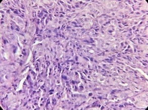
Out of the 31 cases of ulcer, 16 cases were diagnosed as Gastritis and 15 cases were diagnosed as Duodenitis on histopathological examination. Out of the 15 cases of erosion, 8 cases were diagnosed as Gastritis, 3 cases were diagnosed as Duodenitis and 1 case as premalignant on histopathological examination. Out of the 4 cases of polyp, 3 cases were diagnosed as polyp and 1 case as Duodenitis on histopathological examination. Out of the 38 cases of growth, 10 cases were diagnosed as premalignant, 23 cases were diagnosed as Carcinoma Stomach and 4 cases were diagnosed as Carcinoma Duodenum (Table 3) (Figures 1-4).
| Endoscopic findings | Histopathological findings (N=100) | ||||||||
| N= 100 | Normal | Gastritis | Duodenitis | Polyp | Celiac Disease | Metaplasia | Dysplasia | Carinomna Stomach | Carcinoma Duodenum |
| Normal | 6 | 1 | 2 | 0 | 3 | 0 | 0 | 0 | 0 |
| Ulcer | 0 | 16 | 15 | 0 | 0 | 0 | 0 | 0 | 0 |
| Erosion | 0 | 8 | 3 | 0 | 3 | 1 | 0 | 0 | 0 |
| Polyp | 0 | 0 | 1 | 3 | 0 | 0 | 0 | 0 | 0 |
| Growth | 0 | 1 | 0 | 0 | 0 | 1 | 9 | 23 | 4 |
| Total (n=100) | 6 | 26 | 21 | 3 | 6 | 2 | 9 | 23 | 4 |
χ2 value 235.7, p value is <0.0001
Figure 1. Well Differentiated Adenocarcinoma of Stomach .
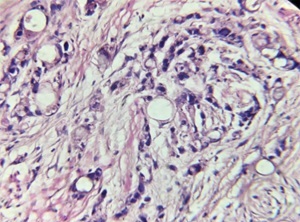
Figure 2. Moderately Differentiated Adenocarcinoma of Stomach .
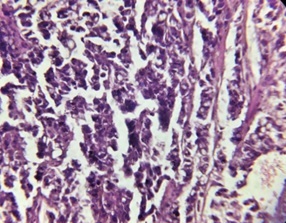
Figure 3. Diffuse Adenocarcinoma with Signet Ring Cells of Stomach .
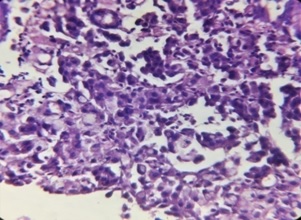
Figure 4. Periampullary Adenocarcinoma of Duodenum .
Out of the 12 normal endoscopic cases, 2 cases were diagnosed as Duodenitis, 1 case as Gastritis on histopathological examination.
When we correlate endoscopic findings with histopathological findings, the association was found to be statistically significant (χ2 value 235.7, p value is <0.0001).
The sensitivity, specificity and positive predictive value of endoscopy and histopathological correlation are 100%, 94.5% and 87.1% respectively. The accuracy is 96%.
Maximum cases (87%) of gastric carcinoma presented as ulcerative growth on endoscopy followed by polypoidal growth (4.3%) and diffuse growth (8.7%) (Table 4).
| Endoscopic Appearance | Total cases (N=23) | Percentage (n=100) |
| Ulcerative | 20 | 87 |
| Polypoid | 1 | 4.30 |
| Diffuse | 2 | 8.70 |
| Nodular | 0 | 0 |
| Total | 23 |
Discussion
The upper GI endoscopy is the diagnostic tool of choice to detect gastroduodenal diseases. Endoscopic examination should always be accompanied by biopsy.Current data suggest that gross visualization alone cannot detect gastric mucosal diseases, only histologic examination can provide accurate diagnosis.In the present study, out of the 100 gastroduodenal biopsies, 59 cases were from gastric region and rest 41 cases were from the duodenum. The present study is consistent with the studies made by Shanmugasamy K et al [5], Vijayabasker Mithun KR et al [6], S. Hirachand et al [7], Deepa Rani et al [8] and Veenaa Venkatesh et al [9] which shows that gastric biopsies are more common compared to duodenal biopsies.
In the present study, the number of male patients were 65 and female patients were 35. Male (M): female (F) ratio is 1.85: 1. The present study is consistent with the studies made by Dr Vishwapriya M. Godkhindi et al [10], Poonam Sharma et al [11] and S. Hirachand et al [7] which shows male preponderance in gastroduodenal lesions.
In the present study, the highest number of cases were in the age group of 41-50 years followed by 51-60 years. The present study is consistent with the studies made by Shanmugasamy K et al [5], Manasa P Kumari et al [12], Vijayabasker Mithun KR et al [6] and Kaur Manpreet et al [13] which show that gastrosuodenal lesions are more common in the 5th and 6th decade of life.
In the present study, the most common clinical complaint was pain abdomen followed by dyspepsia. The present study is consistent with the studies done by Dr Vishwapriya M. Godkhindi et al [10], Rosy Khandelia et al [14] and Sharma S et al [15] which show that pain abdomen is the most common presenting symptom but not consistent with Sonam Pruthi S et al [16] and Shanmugasamy K et al [5] which show dyspepsia as the most common presenting symptom. This disparity may be due to food habits, geographic variations or social habits. In the present study, the most common endoscopic findings was ulcer followed by erosion. The present study was not consistent with the studies made by Sonam Pruthi S et al [16], Manasa P Kumari et al [12] and Kaur Manpreet et al [13] which showed that the most common endoscopic finding was erosion. This disparity may be due to geographical or topographical variation, food habits or socioeconomic factors.
In the present study, out of the 31 cases of ulcer on endoscopy, 16 cases were diagnosed as Gastritis and 15 cases were diagnosed as Duodenitis on histopathological examination. Out of the 15 cases of erosion on endoscopy, 8 cases were diagnosed as Gastritis, 3 cases were diagnosed as Duodenitis and 1 case as premalignant on histopathological examination. Out of the 4 cases of polyp on endoscopy 3 cases were diagnosed as polyp and 1 case as Duodenitis. Out of the 38 cases of growth on endoscopy, 10 cases (26.3%) were diagnosed as premalignant, 23 cases (60.5%) were diagnosed as Carcinoma Stomach and 4 cases (10.5%) were diagnosed as Carcinoma Duodenum. Thus we found a positive correlation between endoscopic and histopathological findings. The present study is consistent with Grace H elta et al [17], Pailoor K et al [18] and Sharma S et al [15] but not consistent with Sharma S et al [19] and Poudel A et al [20]. This disparacy may also be because of geographic and topographical variations, food habits or personal habits.
In the present study, gastric malignancy endoscopically presented most commonly as ulcerative growth followed by polypoidal growth. The present study is consistent with the studies made by Pailoor K et al [18], Kirana Pailoor et al [21], Sharma S et al [19] and Anunayi Jeshtadi et al [22] which show that the most common endoscopic appearance of gastric carcinoma is ulcerative growth.
Strength of the Study
Premalignant lesions are found among study population which show that early detection with the help of endoscopy and biopsy will help in prompt intervention and better management of the patient.
Weakness of the Study
The limitation of the present study is the sample size which is very less and done for a very short duration. The study is a hospital based study due to which follow up of the cases were not possible.
In conclusion, Endoscopy is a less invasive non-surgical procedure. Endoscopic gastroduodenal biopsies help in detecting benign as well as malignant lesions. Endoscopic examination is recommended as the first line of investigation in the work up of a patient with chronic upper abdominal symptoms. Our study showed better correlation with endoscopic findings in cases of malignant lesions but poor correlation in benign lesions with those of histopathological diagnoses. Endoscopic examination alone might miss out in diagnosing majority of the lesions. The combination of endoscopy and histopathological study of gastroduodenal biopsy provide a powerful diagnostic tool for better management of patients.
Acknowledgments
Statement of Transparency and Principals:
· Author declares no conflict of interest
· Study was approved by Research Ethic Committee of author affiliated Institute.
· Study’s data is available upon a reasonable request.
· All authors have contributed to implementation of this research.
References
- Structural organisation and function of Gastrointestinal tract, “http://www.surendranathcollege.org/new/upload/SUMAN_TAMANGStructural%20organisation%20and%20function%20of%20Gastrointestinal%20tract20200402Structural%20organisation%20and%20function%20of%20Gastrointestinal%20tract-converted.pdf”. .
- Peptic-ulcer disease Chan FKL , Leung WK . Lancet (London, England).2002;360(9337). CrossRef
- Internet, https://www.hopkinsmedicine.org/gastroenterology_hepatology/_pdfs/esophagus_stomach/peptic_ulcer_disease.pdf .
- A study on histopathologic spectrum of upper gastrointestinal tract endoscopic biopsies Krishnappa R, Horakerappa MS , Mangala Ali Karar , GouriMangala . Int J Medical Res Health Sciences.2013;2(3):418-24. https://www.nepjol.info/index.php/jbpkihs/article/view/19760/16242.
- “Clinical Correlation of Upper Gastrointestinal Endoscopic Biopsies with Histopathological Findings and To Study the Histopathological Profile of Various Neoplastic and Non-Neoplastic Lesions” http://dx.doi.org/10.20936/jpbms/160226 Shanmugasamy K, Bhavani K, Anandraj Vaithy K, Narashiman R, Dhananjay S Kotasthane . . CrossRef
- “Spectrum of Gastroduodenal Lesions in Endoscopic Biopsies: A Histopathological Study with Endoscopic Correlation”. Available online at http://saspublisher.com/sjams/ Vijayabasker Mithun KR , Rajesh Kumar G, Govindaraj T, Arun Kumar SP . .
- “Histopathological spectrum of upper gastrointestinal endoscopic biopsies” S Hirachand , RR Sthapit , P Gurung , S Pradhanang , R Thapa , M Sedhai , S Regm . JBPKIHS.2018;1(1):68-87.
- A study of morphological spectrum of upper gastrointestinal tract lesions by endoscopy and correlation between endoscopic and histopathological findings Deepa Rani , Stuti Bhuvan , Atul Gupta . Indian Journal of Pathology and Oncology.2019;6(1). CrossRef
- “Histopathological Spectrum of Lesions in Gastrointestinal Endoscopic Biopsies: A Retrospective Study in a Tertiary Care Center in India” Veenaa Venkatesh , Riyana R Thaj . World Journal of Pathology Volume No 10..
- The Histopathological Study Of Various Gastro-duodenal Lesions and Their Association with Helicobacter pylori Infection. Godkhindi V. IOSR Journal of Dental and Medical Sciences.2013;4. CrossRef
- Histopathological Spectrum of various gastroduodenal lesions in North India and prevalence of Helicobacter pylori infection in these lesions: a prospective study Sharma P, Kaul K, Mahajan M, Gupta P. International Journal of Research in Medical Sciences.2015;3. CrossRef
- The Histopathological Study on Helicobacter pylori Associated Gastroduodenal Diseases in a Tertiary Care Hospital, Mysore Kumari M, Sumana MN . 2013;438.
- Correlation between histopathological and endoscopic findings of non-malignant gastrointestinal lesions: an experience of a tertiary care teaching hospital from Northern India Kaur M, Bhasin T, Manjari M, Mannan R, Sharma S, Anand G. Journal of Pathology of Nepal.2018;8. CrossRef
- Histopathologic Spectrum of Upper Gastrointestinal Tract Mucosal Biopsies: A Prospective Study Khandelia R, Saikia M. International Journal of Medical Science and Clinical Invention.2017;4(11). CrossRef
- Clinicopathological Correlation Of Upper Gastrointestinal Tract Endoscopic Biopsies In A Tertiary Care Hospital In Rural Area Of North India Sharma A, Gupta K. International Journal of Advanced Research.2020;8(3). CrossRef
- Evaluation of gastric biopsies in chronic gastritis: Grading of inflammation by Visual Analogue Scale Chakraborti S, Pruthi S, Murali N. Medical Journal of Dr. D.Y. Patil University.2014;7. CrossRef
- A study of the correlation between endoscopic and histological diagnoses in gastroduodenitis Elta GH , Appelman HD , Behler EM , Wilson JA , Nostrant TJ . The American Journal of Gastroenterology.1987;82(8).
- “Histopathological Diagnosis Of Gastric Biopsies In Correlation With Endoscopy – A Study In A Tertiary Care Center” Pailoor K1 , Sarpangala MK2 , Naik RCN . Advance Laboratory Medicine International.2012;3(2):22-31.
- Correlation between Endoscopic and Histopathological Findings in Gastric Lesions Sharma S, Makaju R, Dhakal R, Purbey B. Kathmandu University medical journal (KUMJ).2015;13. CrossRef
- Correlation between Endoscopic and Histopathological Findings in Gastric Lesions Poudel A, Regmi S, Poudel S, Joshi P. Journal of Universal College of Medical Sciences.2013;1. CrossRef
- “Correlation of Endoscopic and Histopathological Diagnosis of Upper Gastrointestinal Lesions: A Study in a Tertiary Care Centre in Coastal Karnataka” Kirana Pailoor , Ramesh Naik C.N,, , Murali Keshava S, Hilda Fernande , Jayaprakash CS , Nisha J. Marla . Indian Journal of Forensic Medicine and Pathology Volume 6 Number 1, January - March 2013..
- “Study Of Gastric Biopsis With Clinicopathological Correlation - A Tertiary Care Centre Experience” Anunayi Jeshtadi , Afzal Moid Mohammad , Madhukar Reddy Kadaru , Ezhil Arasi Nagamuthu , Harika Kalangi , Archana Boddu , Sandeep Kumar Lakkarasu , Ajeta Boila . 2016;3(57):2937-2940.
License

This work is licensed under a Creative Commons Attribution-NonCommercial 4.0 International License.
Copyright
© Asian Pacific Journal of Cancer Biology , 2024
Author Details