Histopathological Findings with Incidental Detection of Prostatic Carcinoma in TURP Specimen: A Retrospective Study in a Tertiary Care Hospital
Download
Abstract
Introduction: In adults prostate weighs upto 20 gms which is a pear shaped organ. Prostatic gland diseases leads to increase morbidity and moratlity in patients. Prostatic adenocarcinoma is one of the common malignancy among men in India. Transurethral resection of prostate (TURP) is one of the common procedures done by the urologist in cases of prostatomegaly.
Aims and Objective: This study was conducted to see various histopathological patterns of prostatic lesions in TURP specimens. Also to evaluate the incidence of incidental adenocarcinoma of prostste.
Materials and Methods: It was a retrospective study done for the duration of 1 year. Total 58 TURP cases were evaluated by retrieving their slides,blocks as well all the clinical informations. All the H & E slides were re examined and new slides were prepared from the paraffin blocks wherever required.
Results: In these study commonest lesion that we encountered in TURP specimen were benign prostatic hyperplasia (89.65%). BPH was associated with prostatitis in 20.69% of cases. Highest BPH cases were seen in the age group of 60-69 years. We also found one case of atypical adenomatoid hyperplasia (AAH) and one case of High grade PIN in our study. We detect 4 cases of incidental adenocarcioma in our study, incidence of which were 6.89 %. Most common age group of adenocarcinoma was 60-69 years. Out of 4 malignant cases 1 case showed perineural invasion in our study.
Conclusion: In TURP specimens, most common lesion are the benign prostatic hyperplasia. However TURP can detect incidental adenocarcinoma also. So it is very important to do routine histopathological examinations of all the TURP specimens to rule out malignancy.
Introduction
In adults prostate weighs upto 20 gms which is a pear shaped organ [1]. Histologically prostatic parenchyma has 4 zones namely peripheral, central, transitional and periurethral zones. Prostatic glands are double layered and lined by basal cuboidal epithelium and secretory columnar epithelium [1]. With increasing age, prostate gland is one of the commonly affected organ in males leading to significant morbidity and mortality. Most common diseases of prostate are Prostatitis, Benign prostatic hyperplasia and carcinoma of prostate. Prostatic enlargement is commonly seen in persons who are above 50 years of age [2]. Prostatis which is defined as inflammation of prostate is associated with dysuria and urinary frequency [3]. BPH is enlargement of prostate which is associated with both stromal and glandular hyperplasia [4]. The incidence of BPH is 75% in 8th decade, 50% in the 5th decade and only 8% in the 4th decade [1]. BPH though not a premalignant lesion, but may be associated with carcinoma prostate in the transition zone [5].
Prostatic carcinoma is one of the common malignancy among men in India and constitute about 5% of all cancers in male [6, 7]. Most important risk factors for occurance of prostatic carcinoma are increasing age,positive family history and high calcium intake [8]. Screening of prostatic lesions constitute estimation of serum PSA level, Digital rectal examination and transrectal ultrasound. However, histopathological examination remains the gold standard for diagnosis. Prior to PSA era, 27% of prostatic carcinoma was detected incidentally by TURP [7].
Aims and Objectives
Our main aim of this study to evaluate the various histomorphological finding of prostatic lesions. This study also tries to know the incidence of incidentally detected prostatic carcinoma.
Materials and Methods
Current study was conducted in the Department of Pathology,Gauhati Medical College and Hospital. It was a retrospective study conducted in the department from September 2022 to August 2023 where all the TURP cases received were analysed. All the relevant histopathological and clinical data of the TURP cases were retrieved and reviewed. All the H & E slides were re examined and new slides were prepared from the paraffin blocks wherever required. Various histopathological patterns were studied in all cases and were classified with reference to age. In case of incidentally detected prostatic carcinoma,grading was done on the basis of Gleason scoring system mentioning the primary and secondary patterns and grade group were assigned.
Inclusion Criteria
All the TURP cases received in the Department of Pathology during the study period.
Exclusion Criteria
All the prostetectomy specimen and prostatic biopsies were excluded from this study.
Results
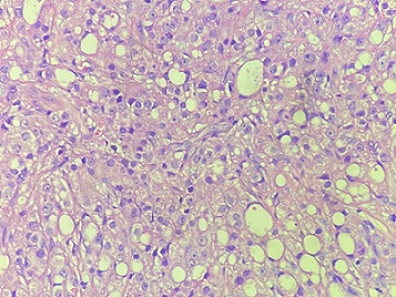
In this study, total 58 cases TURP specimen were examined. Out of 58 cases 40 (68.96%) cases were having BPH (Figure 1 Benign Prostatic Hyperplasia), 12 (20.69%) cases were BPH with prostatitis Figure 2 Benign Prostatic Hyperplasia with Prostatitis), 1 (1.72%) case showed Atypical adenomatoid Hyperplasia, 1 (1.72%) case was High grade PIN (Figure 3 Prostatic Intraepithelial Neoplasia) and 4 (6.89%) cases were detected incidentally as Prostatic Adenocarcinoma (Figure 4 Prostatic adenocarcinoma) (Table 1).
Figure 1. Benign Prostatic Hyperplasia.
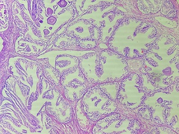
Figure 2. Benign Prostatic Hyperplasia with Prostatitis.
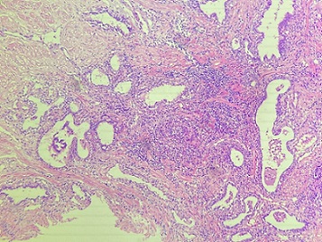
Figure 3. Prostatic Intraepithelial Neoplasia (PIN).
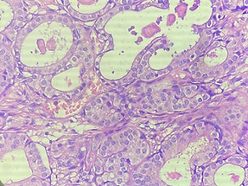
Figure 4. Prostatic Adenocarcinoma (Gleason 5).
| Histopathological diagnosis | Number of cases | Percentage |
| Benign Prostatic hyperplasia (BPH) | 40 | 68.96 |
| BPH with prostatitis | 12 | 20.69 |
| Atypical adenomatoid Hyperplasia (AAA) | 1 | 1.72 |
| PIN Low grade (LGPIN) | 0 | 0 |
| PIN High grade (HGPIN) | 1 | 1.72 |
| Incidental Adenocarcinoma | 4 | 6.89 |
| Total cases | 58 | 100 |
Maximum cases of TURP specimen were from the age group of 60-69 years (Total 30 cases) accounting for 51.72% followed by 14 cases in 50-59 years of age constituting 24.13%. The most common age group presenting with benign lesion was 60-69 years with 28 cases (48.27%) followed by 50-59 years with 14 cases (24.13%). Total 4 incidental adenocarcinoma cases were detected where 2 cases were in the age group of 60-69 (50%) and one each from the age group of 70-79 years and >80 years (Table 2).
| Age (in years) | Benign lesions | Malignant lesions |
| < 50 | 2 | 0 |
| 50-59 | 14 | 0 |
| 60-69 | 28 | 2 |
| 70-79 | 10 | 1 |
| >80 | 4 | 1 |
| Total | 54 | 4 |
Out of total 58 cases 54 were benign lesion and 4 cases were incidentally detected as Prostatic Adenocarcinoma (Table 3).
| Benign lesions | Malignant lesions | Total |
| 54 | 4 | 58 |
| 93.10% | 6.89% | 100% |
We reported 4 cases of adenocarcinoma prostate with modified Gleason Grading system. Most common (50%) Grade Group we found was Grade group 5. Total 2 cases were having grade group 5 out of which one case was having a Gleason score of 9 (4+5) and other case was having a score of 10 (5+5) (Figure 4). One case was having grade group 2 (3+4) and other showed grade group 4 (3+5).
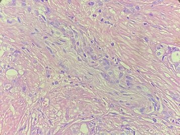
Out of 4 cases one case showed perineural invasion (PNI) (Figure 5) (Table 4).
Figure 5. Perineural Invasion (PNI).
| Grade group | Gleason score | Primary + secondary Pattern | Total cases |
| 1 | 6 | 3+3 | 0 |
| 2 | 7 | 3+4 | 1 |
| 3 | 7 | 4+3 | 0 |
| 4 | 8 | 4+4 | 0 |
| 3+5 | 1 | ||
| 5+3 | 0 | ||
| 5 | 9/10 | 4+5 | 1 |
| 5+4 | 0 | ||
| 5+5 | 1 |
Discussion
In the current study,total 58 cases of TURP specimen were analysed. Benign lesion were found to be common,which is matching with other Indian studies [9-11]. We found 52 (89.65%) BPH cases along with 1 case (1.72%) of PIN and 4 cases (6.89 %) of prostate adenonarcinoma. These findings are similar to previous studies like Sharma A et al, Thapa N et al and Begum Z et al in which benign lesions were found to be common compared to malignant lesions [12-14].
Sharma A et al [14] found 91.02 % of benign cases in their study while Thapa N et al and Begum Z et al found 92.2 % and 96 % respectively of Benign Prostatic hyperplasia cases in their studies [12, 13].
Regarding age group,maximum cases in our study were in the age group of of 60-69 years (Total 30 cases) accounting for 51.72% followed by 14 cases in 50-59 years of age constituting 24.13%.Our findings were matching with other studies like Sharma A et al, Thapa N et al and Shirish C et al [9, 12, 14]. enign prostatic hyperplasia cases were maximum in 7th decade in our current study while some previous studies like Arya RC et al, Kumar M et al., Kasliwal N et al also had the similar observations [15-17].
BPH associated with prostatitis were found in 20.69 % of cases in our study (Total 20 cases out of 58 cases). Sharma et al also found 33.06 % cases prostatitis with BPH [14].
We got 1 case of atypical adenomatoid hyperplasia in our study. Atypical adenomatoid hyperplasia (AAH) can mimick adenocarcinoma of prostste [18] and usually involves the transitional zone. Incidence of AAH is 1.72 % in our study. Sharma et al (1.22), Garg et al (1.65%) and Puttaswamy K et al (2%) also had the similar findings in their respective studies [7, 14, 19].
We report one case of High grade PIN in our study, low grade PIN was not reported in our study.The incidence of PIN was found to be 1.72% in our study. Incidence was PIN was found to be s 2.3 - 5.5% in some similar studies like Brawn PN et al, Alsaikafi NF et al, Gaudin PB et al [20-22].
There were 4 cases of incidentally detected adenocarcioma in our study. The incidence of incidental adenocarcinoma was 6.89 %. Sharma et al found an incidence of 3.26 % in their study [14] while V Sailaja et al found an incidence of 8.57 % in their studies [23]. Yadav et.al also had a similar finding in their study where they got an incidence of 7.00% of prostatic adenocarcinoma [24]. Two cases were having grade group 5 (Gleason score of 9 and 10) while 2 cases were in the grade group of 2 (Gleason score 7) and 4 (Gleason score 8). It has been seen that post PSA era, incidence of incidental detection of adenocarcinoma of prostate has decreased. However incidental detection of prostatic adenocarcinoma is not uncommon now a days also.
In conclusion, from this current study we concluded that in TURP specimens most common lesion are the benign prostatic hyperplasia.Many cases of prostatitis were also associated with BPH. Commonest age group involved by BPH were found to be the 7th decade. We also concluded that incidental detection of proststic adenocarcinoma is not uncommon. Though PSA is a good marker for adenocarcinoma of prostate, still few cases were clinically thought to be BPH only which came out to be malignant in TURP histopathology assessment.
Conflict of Interests
The authors declare that there is no conflict of interests regarding the publication of this paper.
References
- Rosai and Ackerman’s surgical pathology tenth edition ;:12-87.
- The lower urinary tract and male genital system Epstein JI . Robbins and Cotran Pathologic Basis of Diseases.2005.
- Prostate. http://www.srhmatters.org/sexualhealth/prostate/ .
- Nonneoplastic lesions of the prostate and bladder Harik LR , O'Toole KM . Archives of Pathology & Laboratory Medicine.2012;136(7). CrossRef
- Decision-making strategies for patients with localized prostate cancer Diefenbach MA , Dorsey J, Uzzo RG , Hanks GE , Greenberg RE , Horwitz E, Newton F, Engstrom PF . Seminars in Urologic Oncology.2002;20(1). CrossRef
- Consolidated Report of Population Based Cancer Registries 2001-2004: Incidence and Distribution of Cancer. Bangalore (IND): Coordinating Unit, National Cancer Registry Programme, Indian Council of Medical Research Egevad L . 2006.
- Histopathological spectrum of 364 prostatic specimens including immunohistochemistry with special reference to grey zone lesions Garg M, Kaur G, Malhotra V, Garg R. Prostate International.2013;1(4). CrossRef
- Prostate Cancer Risk Factors Prostate cancer foundation.2001.
- Clinico-pathological study of benign and malignant lesions of prostate Shirish C, Jadhav PS , Anwekar SC , Kumar H, Buch AC , Chaudhari US . IJPBS.2013;3:162-178.
- The histomorphological study of prostate lesions Joshee A, Sharma KCL . IOSR-JDMS.2015;14:85-89.
- Prostate biopsies: a five year study at a tertiary care centre Burdak P, Joshi N, Nag BP , Jaiswal RM . IJSR.2015;4:420-3.
- Incidence of Carcinoma Prostate in Transurethral Resection Specimen in a Teaching Hospital of Nepal Thapa N, Shris S, Pokharel N, Tambay Y, Kher Y, Acharya S. Journal of Lumbini Medical College.2016;4. CrossRef
- Study of Various Histopathological Patterns in Turp Specimens and Incidental Detection of Carcinoma Prostate Begum Z, Attar A, Tengli M, Ahmed M. Indian Journal of Pathology and Oncology.2015;2. CrossRef
- Histomorphological spectrum of prostatic lesions: a retrospective analysis of transurethral resection of prostate specimens Sharma A, Gandhi S, Khajuria A, Goswami K. International Journal of Research in Medical Sciences.2017. CrossRef
- Pattern of prostatic lesions in Chhattisgarh Institute of Medical Sciences, Bilaspur: a retrospective tertiary hospital based study Arya RC , Minj MK , Tiwari AK , Bhardwaj A, Singh D, Deshkar AM . Int J Sci Stud.2015;3:179-82.
- Clinicopathological study of prostate lesions Kumar M, Khatri SL , Saxena V, Vijay S. IJBAMR.2016;6:695-704.
- Pattern of prostatic disease- a histopathological study with clinical correlation Kasliwal N. EJPMR.2016;3(3):589-97.
- Pseudoneoplastic mimics of prostate and bladder carcinomas Hameed O, Humphrey PA . Archives of Pathology & Laboratory Medicine.2010;134(3). CrossRef
- Histopathological Study of Prostatic Biopsies in Men with Prostatism Puttaswamy K, Parthiban R, Shariff S. Journal of Medical Sciences and Health.2018;2. CrossRef
- Histologic grading study of prostate adenocarcinoma: The development of a new system and comparison with other methods—A preliminary study Brawn PN , Ayala AG , Eschenbach ACV , Hussey DH , Johnson DE . Cancer.1982;49(3). CrossRef
- High-grade prostatic intraepithelial neoplasia with adjacent atypia is associated with a higher incidence of cancer on subsequent needle biopsy than high-grade prostatic intraepithelial neoplasia alone Alsikafi N. F., Brendler C. B., Gerber G. S., Yang X. J.. Urology.2001;57(2). CrossRef
- Incidence and clinical significance of high-grade prostatic intraepithelial neoplasia in TURP specimens Gaudin P. B., Sesterhenn I. A., Wojno K. J., Mostofi F. K., Epstein J. I.. Urology.1997;49(4). CrossRef
- Sailaja V, Jyothi C, Sreedhar VV , Rao MN , Ram PS . Saudi J Pathol Microbiol.(SJPM) ISSN 2518-3362 (Print). .2018;3(8):221-225.
- Study of various histopathological patterns in prostate biopsy Yadav M, Desai H, Goswami H. Int J Cur Res Rev.2017;9(21):58-63.
License

This work is licensed under a Creative Commons Attribution-NonCommercial 4.0 International License.
Copyright
© Asian Pacific Journal of Cancer Biology , 2025
Author Details