Histopathological Study of Gastroduodenal Biopsies and to See the Prevalence of Helicobacter Pylori with the Help of Special Stains in Selected Cases in North East India- A Cross Sectional Study in a Tertiary Care Centre
Download
Abstract
Background: Acid peptic disease (APD), including gastric ulcers, duodenal ulcers, and gastro esophageal reflux disease is a common disorder of the gastrointestinal region, the pathogenesis of which involves an imbalance between acid secretion and gastric mucosal defenses. Acid peptic diseases mostly affect the esophagus, stomach, and duodenum. The imbalance could be caused by factors such as H pylori infection and acid secretory abnormalities in APD. Helicobacter pylori infection is a factor in 85% to 100% of duodenal ulcers and 70% to 90% of gastric ulcers. Besides, malignancies are also fairly common in these regions. Due to long standing APD or H. Pylori infection it may lead to dysplastic changes or malignancy in this region. These disorders are easily detected by endoscopy nowadays. This is a cross-sectional study done in 100 cases of gastroduodenal biopsies in a 1 year period.
Aims and Objectives: To see the histopathological spectrum of gastroduodenal lesions and to detect the presence of H. pylori infection in suspected cases with the help of special stains in North East India.
Result: There were 13 cases (%) of Helicobacter pylori gastritis diagnosed by H&E and Giemsa staining procedure. Out of the 13 cases, the highest number of cases, 46.1% was in the age group of 51-60 years. Out of the 13 cases of H. Pylori gastritis, 11 cases (84.6%) were male and 2 cases (15.3%) were female. The male: female ratio is 5.5 : 1.
Introduction
Gastroduodenal disorders are among the common clinical disease, out of which the inflammatory and neoplastic lesions are particularly common [1]. The most common abnormalities which are frequently reported by endoscopy are duodenal ulcer (2.3-12.7%), gastric ulcer (1.6-8.2%) and gastric malignancy (0-3.4%) [2]. Helicobacter pylori is one of the causes of peptic ulcer. H.pylori is responsible for more than 90% of duodenal ulcers and 65% of gastric ulcers [3, 4]. As H. Pylori infection is treatable, it is of utmost importance that it should be detected at the earliest [5]. H. Pylori have been linked to various disease conditions like acute and chronic gastritis, non ulcer dyspepsia, gastric adenocarcinoma and gastric Non-Hodgkin’s Lymphoma of mucosa associated lymphoid tissue (MALT). The annual incidence of H pylori in the developed country and developing country are 0.3-0.7 % and 6-14% respectively [6]. The International Agency for research on cancer (IARC), sponsored by WHO in the year 1994 categorised H. Pylori as a class 1 carcinogen and a definite cause of gastric carcinoma in humans [7]. The most common cancer of stomach worldwide is gastric adenocarcinoma, early detection of malignancy thus improves the survival rate of the patients [8].
Aim of the Study
To see the histopathological spectrum of gastroduodenal lesions and to detect the presence of H. pylori infection in suspected cases with the help of special stains in North East India.
Materials and Methods
This is a hospital based cross sectional study which has been carried out in the Department of Pathology in collaboration with the department of Gastroenterology, Gauhati Medical College and Hospital for a period of 1 year from July 2020 to June 2021. A total of 100 patients with chronic upper abdominal symptoms are selected for the study. All endoscopic gastroduodenal biopsies are received in our department in 10% neutral buffered formalin solution. The tissue is then processed, sections are made of 3 micrometre thickness and then stained with Haematoxylin and Eosin stain. Special stain is then done with Giemsa stain for H. pylori identification in selected cases.
SPECIAL (GIEMSA) STAINING PROCEDURE
1. Bring sections to water
2. Giemsa stain- 5 mins
3. Quick dehydration in alcohol
4. Clearing in Xylene
5. Mount in DPX
Result- H. pylori is stained dark blue, background is stained pink to dark blue
Exclusion Criteria
1. Biopsy done for therapeutic purpose
2. Cases where biopsy cannot be done
3. Cases where consent not given
4. Autolysed specimen.
Ethical Clearance
The study has been ethically approved by the Institutional ethical committee (letter no- 190/2007/ dt-11/dec-2019/61). The patients were explained about the research project and written consent was taken from patients or guardians in case of minors.
Results
A total number of 100 patients who underwent gastroduodenal biopsies and fulfilled the inclusion criteria as per stated were enrolled during the study. Depending upon the symptoms and clinical presentation, gastric biopsies were taken from 59 cases and duodenal biopsies from 41 cases. The male: female, M: F ratio is 1.85:1. The age of the patients varied from 25 to 84 years with peak incidence in the 5th decade. Abdominal pain was the most common clinical complaint (Table 1).
| Age range | Total | Percentage |
| 21-30 | 8 | 8 |
| 31-40 | 19 | 19 |
| 41-50 | 35 | 35 |
| 51-60 | 26 | 26 |
| 61-70 | 8 | 8 |
| 71-80 | 3 | 3 |
| 81-90 | 1 | 1 |
| Total (N=100) | 100 |
Out of the 100 gastroduodenal biopsy studied, 56% were diagnosed as benign, 11% as premalignant condition and 27% as carcinoma (Table 2).
| Categories | Type of lesion | No of cases | Total cases | Percentage (N=100) |
| N=100 | ||||
| Normal | Normal findings | 6 | 6 | 6 |
| Benign | Gastritis | 26 | 56 | 56 |
| Duodenitis | 21 | |||
| Polyp | 3 | |||
| Celiac Disease | 6 | |||
| Premalignant | Gastric Intestinal Metaplasia | 2 | 11 | 11 |
| Low Grade Dysplasia | 8 | |||
| High Grade Dysplasia | 1 | |||
| Malignant | Carcinoma Stomach | 23 | 27 | 27 |
| Carcinoma Duodenum | 4 |
In the present study, benign category had the highest number of cases, maximum of which belonged to the age group of 41-50 years. Among the benign category maximum numbers of cases (46%) were diagnosed as gastritis (Table 3).
| Age | Histopathological Diagnosis | ||||||
| group | Gastritis | Duodenitis | Polyp | Celiac Disease | Gastric Intestinal Metaplasia | Dysplasia | Carcinoma |
| 21-30 | 3 | 2 | 0 | 2 | 0 | 0 | 1 |
| 31-40 | 7 | 3 | 0 | 1 | 1 | 1 | 4 |
| 41-50 | 6 | 13 | 2 | 2 | 1 | 4 | 4 |
| 51-60 | 8 | 2 | 1 | 1 | 0 | 2 | 11 |
| 61-70 | 2 | 1 | 0 | 0 | 0 | 2 | 3 |
| 71-80 | 0 | 0 | 0 | 0 | 0 | 0 | 3 |
| 81-90 | 0 | 0 | 0 | 0 | 0 | 0 | 1 |
| Total (N=94) | 26 | 21 | 3 | 6 | 2 | 9 | 27 |
Maximum number of cases from the premalignant category belonged to age group 41-50 years. Maximum number of malignant cases belonged to the age group 51-60 years.
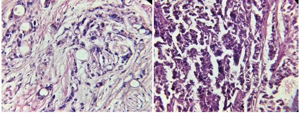
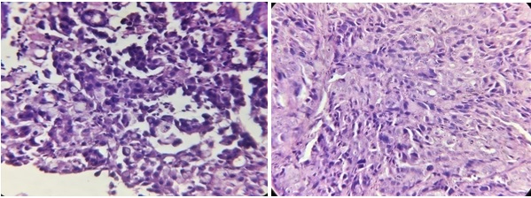
27 cases were diagnosed as carcinoma, out of which 23 cases (85.1%) are Carcinoma Stomach and 4 cases (14.9%) are Carcinoma Duodenum. The age group 51-60 had the maximum number of cases (Figure 1 and 2).
Figure 1. a) Well Differentiated Adenocarcinoma of stomach. b) Moderately differentiated Adenocarcinoma of stomach.

Figure 2. a) Diffuse Adenocarcinoma with Signet Ring Cells of Stomach, b) Periampullary adenocarcinoma of duodenum.

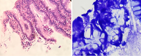
On the basis of endoscopic and histopathological findings, 85 cases (26 gastritis, 21 duodenitis, 11 premalignant and 27 malignant) have been considered for special stain (Giemsa stain) technique to observe the presence or absence of H. pylori in the biopsy tissue. And the result of the special staining technique are as follows: Out of the 13 Helicobacter pylori positive cases diagnosed by Giemsa staining procedure, the highest number (38.4%.) of cases belonged to the age group of 51-60 years (Table 4) (Figure 3).
| Age | Helicobacter Pylori | Total cases (N=85) | |
| Positive | Negative | ||
| 21-30 | 0 | 6 | 6 |
| 31-40 | 3 | 13 | 16 |
| 41-50 | 3 | 25 | 28 |
| 51-60 | 5 | 18 | 23 |
| 61-70 | 2 | 6 | 8 |
| 71-80 | 0 | 3 | 3 |
| 81-90 | 0 | 1 | 1 |
| Total | 13 | 72 | 85 |
Figure 3. a) H and E Stain Showing H. pylori in Gastric Mucosa, b) Giemsa stain showing H. pylori in gastric mucosa.

Out of 13 Helicobacter pylori cases, 53.8% were biopsy of antrum and 23.1% were of pyloric region. 7.7% each were from D1 and D2 region (Table 5).
| Site of gastroduodenal biopsies | H. pylori positive cases |
| Antrum | 8 (61.5%) |
| Pylorus | 3 (23.1%) |
| D1 region | 1 (7.7%) |
| D2 region | 1 (7.7%) |
13 cases were found to be H. pylori positive whereas rest 72 cases were found to be H. pylori negative (Table 6).
| Sex | Helicobacter pylori | Total (N=85) | |
| Positive | Negative | ||
| Male | 11 | 47 | 58 |
| Female | 2 | 25 | 27 |
| Total | 13 | 72 | 85 |
Chi- square value is 22.05 and p value <0.0001, which is statistically highly significant (Table 7).
| Histopathological findings | No of H. pylori positive cases | No of H. pylori negative cases | Total (N=85) |
| Gastritis | 11 | 15 | 26 |
| Duodenitis | 2 | 19 | 21 |
| Premalignant | 0 | 11 | 11 |
| Malignant | 0 | 27 | 27 |
| Total (N=85) | 13 | 72 | |
| χ2 value 22.05, p value <0.0001 |
When we correlate the findings of H&E stain and Giemsa stain, the sensitivity, specificity and positive predictive value are 100% respectively.
Discussion
The most common cause of peptic ulcer not associated with NSAIDS use is H pylori infection. Almost all gastric ulcers occur on the lesser curvature in the antrum, in an area close to the incisura angularis. Gastric ulcers which occur in the greater curvature and in other areas of the stomach such as the fundus are more likely to be associated with chronic NSAIDS use than with H. Pylori. Histological examination has been used as a diagnostic tool for H pylori [9]. Various staining procedures are used for the detection of H pylori which includes H&E, Giemsa, Gram’s stain, 1% methylene blue, Warthin Starry silver stain and fluorescent stains [10].
Giemsa stain is named after Gustav Giemsa. The staining method was designed primarily for the detection of malarial parasites. But it was also employed in histology because of high quality staining of the chromatin and nuclear membrane and different qualities of cytoplasmic staining depending on the cell type. Kiel classification also used this stain for classifying lymphoma [11]. The modified Giemsa stain is a reliable, cheap, easy to perform and convenient procedure for diagnosing H pylori in gastric biopsy specimens [12].
In the present study, out of the 100 gastroduodenal biopsies, 59 cases were from gastric region and rest 41 cases were from the duodenum. The present study is consistent with the studies made by Shanmugasamy K et al [13], Vijayabasker Mithun KR et al [14], S. Hirachand et al [15], Deepa Rani et al [16] and Veenaa Venkatesh et al [17] which shows that gastric biopsies are more common compared to duodenal biopsies.
In the present study, the number of male patients were 65 and female patients were 35. Male (M): female (F) ratio is 1.85 : 1. The present study is consistent with the studies made by Dr Vishwapriya M. Godkhindi et al [18], Poonam Sharma et al [19] and S. Hirachand et al [15] which shows male preponderance in gastroduodenal lesions.
In the present study, the highest number of case were in the age group of 41-50 years followed by 51-60 years. The present study is consistent with the studies made by Shanmugasamy K et al [13], Manasa P Kumari et al [20], Vijayabasker Mithun KR et al [14] and Kaur Manpreet et al [21] which show that gastrosuodenal lesions are more common in the 5th and 6th decade of life.
In the present study, the most common clinical complaint was pain abdomen followed by dyspepsia. The present study is consistent with the studies done by Dr Vishwapriya M. Godkhindi et al [18], Rosy Khandelia et al [22] and Sharma S et al [23] which show that pain abdomen is the most common presenting symptom but not consistent with Sonam Pruthi S et al [24] and Shanmugasamy K et al [13] which show dyspepsia as the most common presenting symptom. This disparity may be due to food habits, geographic variations or social habits.
In the present study, there were 56 non neoplastic cases, 11 premalignant cases and 27 malignant cases. The present study is consistent with the studies done by S. Hirachand et al [15], Kaur Manpreet et al [21], Deepa Rani et al [16] and Sharma S et al [23] which show non neoplastic lesions are more common than neoplastic lesions in the gastroduodenal region.
In the present study, there were 13 cases (15.3%) of Helicobacter pylori gastritis diagnosed by H & E and Giemsa staining procedure. Out of the 13 cases, the highest number of cases was in the age group of 51-60 years. The present study is consistent with Deepa Rani et al [16], Rupendra Thapa et al [25] and Luiz Carlos Bertges et al [26] but not consistent with Shanmugasamy K et al [13] and Anunayi Jeshtadi et al [27]. This disparity may also be because of geographic variations, food habits or socioeconomic habits.
Strength of the Study
Premalignant lesions are found among study population which show that early detection with the help of endoscopy and biopsy will help in prompt intervention and better management of the patient. Also detection of H. Pylori in both H&E and Giemsa stain helps in its proper management and treatment.
Weakness of the Study
The limitation of the present study is the sample size which is very less and done for a very short duration. The study is a hospital based cross sectional study due to which follow up of the cases were not possible. Also silver staining for H.Pylori is better diagnostic method but since not available n our department so we couldnot do it.
In conclusion, application of Giemsa stain in gastroduodenal biopsy for detection of H. pylori may predict the biological behavior of the lesion. The combination of endoscopy and histopathological study of gastroduodenal biopsy provide a powerful diagnostic tool for better management of patients.
Acknowledgments
Statement of Transparency and Principals:
· Author declares no conflict of interest
· Study was approved by Research Ethic Committee of author affiliated Institute .
· Study’s data is available upon a reasonable request.
· All authors have contributed to implementation of this research.
References
- Helicobacter pylori in gastroduodenal diseases. Lawal OO , Rotimi O, Okeke I. Journal of the National Medical Association.2007;99(1).
- Role of endoscopy and biopsy in the work up of dyspepsia Tytgat G. N. J.. Gut.2002;50 Suppl 4(Suppl 4). CrossRef
- Campylobacter pylori and Peptic Ulcer Disease Graham DY . Gastroenterology.1989;96(2, Part 2). CrossRef
- Helicobacter pylori and ulcerogenesis Peura D. A.. The American Journal of Medicine.1996;100(5A). CrossRef
- Comparison of endoscopic brush cytology with biopsy for detection of Helicobacter pylori in patients with gastroduodenal diseases Ahluwalia C., Jain M., Mehta G., Kumar N.. Indian Journal of Pathology & Microbiology.2001;44(3).
- Comparative Evaluation of the Diagnostic Tests for Helicobcter Pylori and Dietary Influence for its Acuisition in Dyspeptic Patients: A Rural Hospital Based Study in Central India Kaore NM , Ngdeo NV , Thombare VR . Journal of Clinical and Diagnostic Research.2012;6(4):636-41.
- Clinical Presentation, Histological Findings and Prevalence of Helicobacter pylori in Patients of Gastric Carcinoma Kabir Ma, Barua Rinti, Masud H, Ahmed Ds, Islam M. M., Karim E, Sarker Mn, Barman Rc. Faridpur Medical College Journal.2011;6. CrossRef
- Histopathological study of gastric carcinoma with associated precursor lesions Manasa G, Manjunath G. Indian Journal of Pathology and Oncology.2016;3. CrossRef
- Robbins and Cotran Pathologic Basis of Disease. 8th ed. Philadelphia: Saunders Elsevier Inc Kumar V , Abbas AK , Fausto N , Aster JC , editors . 2010;:776-779.
- Helicobacter pylori Infection in Endoscopic Biopsy Specimens of Gastric Antrum: Laboratory Diagnosis and Comparative Efficacy of Three Diagnostic Tests Malik G. M., Mubarik M., Kadla S. A.. Diagnostic and Therapeutic Endoscopy.1999;6(1). CrossRef
- The Giemsa stain: its history and applications Barcia JJ . International Journal of Surgical Pathology.2007;15(3). CrossRef
- Histological identification of Helicobacter pylori: comparison of staining methods Rotimi O., Cairns A., Gray S., Moayyedi P., Dixon M. F.. Journal of Clinical Pathology.2000;53(10). CrossRef
- Clinical Correlation of Upper Gastrointestinal Endoscopic Biopsies with Histopathological Findings and To Study the Histopathological Profile of Various Neoplastic and Non-Neoplastic Lesions Shanmugasamy K, Bhavani K, Anandraj Vaithy K, Narashiman R, Dhananjay S Kotasthane . . CrossRef
- “Spectrum of Gastroduodenal Lesions in Endoscopic Biopsies: A Histopathological Study with Endoscopic Correlation” Vijayabasker Mithun KR , Rajesh Kumar G, Govindaraj T, Arun Kumar SP . Available online at http://saspublisher.com/sjams/..
- “Histopathological spectrum of upper gastrointestinal endoscopic biopsies” Hirachand S, Sthapit PR , Gurung P, Pradhanang S, Thapa R, Sedhai M, Regmi S. JBPKIHS.2018;1(1):68-67.
- A study of morphological spectrum of upper gastrointestinal tract lesions by endoscopy and correlation between endoscopic and histopathological findings Deepa Rani , Stuti Bhuvan , Atul Gupta . Indian Journal of Pathology and Oncology.2019;6(1). CrossRef
- Histopathological Spectrum of Lesions in Gastrointestinal Endoscopic Biopsies: A Retrospective Study in a Tertiary Care Center in India Veenaa Venkatesh , Riyana R Thaj . World Journal of Pathology.;(10).
- The Histopathological Study Of Various Gastro-duodenal Lesions and Their Association with Helicobacter pylori Infection. Godkhindi V. IOSR Journal of Dental and Medical Sciences.2013;4. CrossRef
- Histopathological Spectrum of various gastroduodenal lesions in North India and prevalence of Helicobacter pylori infection in these lesions: a prospective study Sharma P, Kaul K, Mahajan M, Gupta P. International Journal of Research in Medical Sciences.2015;3. CrossRef
- The Histopathological Study on Helicobacter pylori Associated Gastroduodenal Diseases in a Tertiary Care Hospital Manasa P Kumari , Sunila , Sumana MN , Nandeesh H. 2015. www.ijsr.net;4(7).
- Correlation between histopathological and endoscopic findings of non-malignant gastrointestinal lesions: an experience of a tertiary care teaching hospital from Northern India Kaur M, Bhasin T, Manjari M, Mannan R, Sharma S, Anand G. Journal of Pathology of Nepal.2018;8. CrossRef
- Histopathologic Spectrum of Upper Gastrointestinal Tract Mucosal Biopsies: A Prospective Study Khandelia R. International Journal Of Medical Science And Clinical Invention.2017;4. CrossRef
- Correlation between Endoscopic and Histopathological Findings in Gastric Lesions Sharma S, Makaju R, Dhakal R, Purbey B. Kathmandu University medical journal (KUMJ).2015;13. CrossRef
- Evaluation of gastric biopsies in chronic gastritis: Grading of inflammation by Visual Analogue Scale Chakraborti S, Pruthi S, Murali N. Medical Journal of Dr. D.Y. Patil University.2014;7. CrossRef
- Histopathological Study of Endoscopic Biopsies Thapa R, Lakhey M, Yadav P, Kandel P, Aryal C, Subba K. JNMA; journal of the Nepal Medical Association.2013;52. CrossRef
- Comparison between the endoscopic findings and the histological diagnosis of antral gastrites Bertges L, Dibai F, Bezerra G, Oliveira E, Aarestrup F, Bertges K. Arquivos de Gastroenterologia.2018;55. CrossRef
- Study Of Gastric Biopsis With Clinicopathological Correlation - A Tertiary Care Centre Experience Jeshtadi A, Mohammad A, Kadaru M, Nagamuthu E, Kalangi H, Boddu A, Lakkarasu S, Boila A. Journal of Evidence Based Medicine and Healthcare.2016;3. CrossRef
License

This work is licensed under a Creative Commons Attribution-NonCommercial 4.0 International License.
Copyright
© Asian Pacific Journal of Cancer Biology , 2024
Author Details