VEGF as a Predictor of Mitotic Activity and Brain Invasion in Meningioma
Download
Abstract
Background: Meningiomas exhibit variable biological behavior, with mitotic activity and brain invasion being critical histopathological markers of aggressiveness. While Vascular Endothelial Growth Factor (VEGF) is implicated in tumor progression, its precise role in driving these specific aggressive features in meningioma remains to be fully elucidated. This study investigated the association between immunohistochemical VEGF expression, mitotic activity, and brain invasion in meningioma.
Methods: We conducted a retrospective, cross-sectional analysis of 73 surgically resected meningioma specimens. VEGF expression was semi-quantitatively scored via immunohistochemistry (negative, weak, moderate, strong) and correlated with mitotic counts (per 10 high-power fields, HPF) and the presence of brain invasion. Associations were assessed using the chi-square test.
Results: High VEGF expression was significantly associated with a higher mitotic index (≥4 mitoses/10 HPF) (p=0.005); notably, 80% (8/10) of cases with high mitotic activity demonstrated moderate to strong VEGF expression. Furthermore, VEGF expression was significantly elevated in brain-invasive meningiomas compared to their non-invasive counterparts (p=0.007), with 75% (3/4) of invasive cases exhibiting strong VEGF expression.
Conclusion: Elevated VEGF expression strongly and statistically significantly correlates with both increased mitotic activity and brain invasion in meningiomas. These findings underscore VEGF’s crucial role as a mediator of tumor proliferation and parenchymal infiltration, positioning it as a potential biomarker for aggressive meningioma and a promising target for future therapeutic interventions.
Introduction
Meningiomas are the most common primary intracranial tumors, comprising approximately 37% of all central nervous system (CNS) neoplasms [1]. While the majority are benign (World Health Organization [WHO] Grade I) and indolent, a significant subset demonstrates aggressive behavior, posing substantial clinical challenges due to higher rates of recurrence and resistance to adjuvant therapies [2].
The 2021 WHO classification system stratifies meningiomas into three grades based on histopathological features. Grade II (atypical) and Grade III (anaplastic) tumors are defined by criteria including elevated mitotic activity (≥4 mitoses/10 high-power fields, HPF) and/ or the presence of brain invasion [3]. Brain invasion, characterized by tumor cells breaching the pial-glial barrier to infiltrate the brain parenchyma, is a definitive criterion for Grade II designation and is strongly associated with recurrence [4]. Despite their prognostic importance, the molecular drivers underpinning these aggressive phenotypes remain incompletely understood. Tumor growth and invasion are fundamentally dependent on angiogenesis, a process regulated by key cytokines such as Vascular Endothelial Growth Factor (VEGF) [5]. VEGF promotes endothelial cell proliferation and vascular permeability, and its overexpression is linked to aggressive behavior and poor prognosis in numerous CNS malignancies, including gliomas and brain metastases [6, 7]. While prior studies have associated VEGF with higher meningioma grades and increased vascularity [8, 9], a direct and specific investigation of its relationship with the core histological drivers of grade mitotic activity and brain invasion is warranted.
Therefore, this study aims to elucidate the association between immunohistochemical VEGF expression and these two critical markers of meningioma aggressiveness. We hypothesize that elevated VEGF expression correlates with both a higher mitotic index and the presence of brain invasion, suggesting its role in promoting tumor proliferation and infiltration.
Materials and Methods
Study Design and Patient Cohort
This cross-sectional analytical study was conducted on 73 archived meningioma specimens obtained from patients who underwent surgical resection at Wahidin Sudirohusodo General Hospital, Makassar, Indonesia, between January 2023 and June 2024. Inclusion criteria were a histopathological diagnosis of meningioma based on the 2021 WHO criteria. Patients who received preoperative radiotherapy or embolization were excluded. This study received approval from the Hasanuddin University institutional ethics committee.
Histopathology and Immunohistochemistry
Formalin-fixed, paraffin-embedded (FFPE) blocks were sectioned at 4-μm thickness. One section was stained with hematoxylin and eosin (H&E) for histopathological assessment. Mitotic activity was counted in 10 HPF in the most mitotically active areas. Brain invasion was defined as tumor cell infiltration beyond the pia-glial barrier into the brain parenchyma.
For immunohistochemistry (IHC), sections were stained with a primary rabbit polyclonal anti-human VEGF-A antibody (GTX102643; GeneTex, Irvine, CA). Staining was performed using an avidin-biotin-peroxidase complex technique with a hematoxylin counterstain.
VEGF Expression Scoring
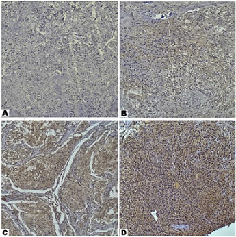
VEGF immunoreactivity in the cytoplasm and membrane of tumor cells was evaluated by two independent pathologists blinded to the clinical data, using a light microscope (Olympus CX43). Expression was scored using a semi-quantitative system based on staining intensity and the percentage of positive cells, adapted from previous studies [10, 11] (representative examples are shown in Figure 1).
Figure 1. Characteristics of VEGF Expression in Meningioma. Representative Immunohistochemical Staining Showing VEGF Expression Levels in Meningioma Cases: (A) negative expression; (B) low expression; (C) moderate expression; and (D) strong expression (100× magnification).

The scoring was categorized as follows: 0 (Negative):
no staining; 1 (Weak): faint staining in <10% of tumor cells; 2 (Moderate): weak-to-moderate staining in 10-50% of cells; and 3 (Strong): moderate-to-strong staining in>50% of cells.
Statistical Analysis
Data were analyzed using SPSS for Windows (Version 29.0). Descriptive statistics were used for clinicopathological characteristics. The association between categorical variables (VEGF expression level vs. mitotic activity category; VEGF expression vs. brain invasion) was analyzed using the Chi-square test. A p-value < 0.05 was considered statistically significant.
Results
Clinicopathological Characteristics
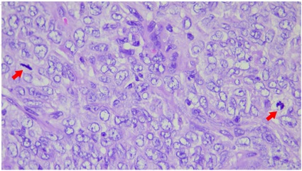
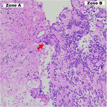
A total of 73 meningioma cases were included. The cohort was predominantly female (n=64, 87.7%) with a mean age of 49 years. The most common tumor location was the convexity (n=31, 42.5%), and the most frequent histopathological subtype was meningothelial (n=48, 65.8%). In accordance with the 2021 WHO classification, 60 cases (82.2%) were Grade I, 11 (15.1%) were Grade II, and 2 (2.7%) were Grade III. High mitotic activity was observed in the higher-grade tumors (Figure 2), and brain invasion was identified in 4 cases (5.5%) (Figure 3).
Figure 2. Mitotic Activity in a Grade III Anaplastic Meningioma Sample. Histopathologic image showing mitotic activity (indicated by red arrow) in a Grade III anaplastic meningioma (Hematoxylin and Eosin stain, 400× magnification).

Figure 3. Histopathological Illustration of Brain Invasion by Meningioma. The red arrow indicates brain invasion by meningioma tissue. Two distinct zones are visible: Zone A shows the brain parenchyma, while Zone B shows the invading meningioma tissue protruding into it (Hematoxylin and Eosin stain, 200× magnification).

Detailed clinicopathological data are presented in Table 1.
| Characteristics | Total (N=73) | n (%) |
| Sex | ||
| Female | 64 | 87.7 |
| Male | 9 | 12.3 |
| Age Group (years) | ||
| <21 | 1 | 1.4 |
| 21-30 | 2 | 2.7 |
| 31-40 | 11 | 15.1 |
| 41-50 | 26 | 35.6 |
| 51-60 | 24 | 32.9 |
| 61-70 | 3 | 4.1 |
| >70 | 6 | 8.2 |
| Tumor Location | ||
| Convexity | 31 | 42.5 |
| Sphenoid wing | 12 | 16.4 |
| Parasagittal | 7 | 9.6 |
| Olfactory groove | 5 | 6.8 |
| Suprasellar | 4 | 5.5 |
| Falx | 3 | 4.1 |
| Multiple | 2 | 2.7 |
| Optic nerve sheath | 2 | 2.7 |
| Retrobulbar | 2 | 2.7 |
| Spheno-orbital | 2 | 2.7 |
| Fossa posterior | 1 | 1.4 |
| Cerebellopontine angle (CPA) | 1 | 1.4 |
| Petroclival | 1 | 1.4 |
| Histopathological Subtype | ||
| Meningothelial | 48 | 65.8 |
| Fibrous | 7 | 9.6 |
| Microcystic | 2 | 2.7 |
| Transitional | 2 | 2.7 |
| Angiomatous | 1 | 1.4 |
| Psammomatous | 1 | 1.4 |
| Atypical | 7 | 9.6 |
| Clear cell | 2 | 2.7 |
| Chordoid | 1 | 1.4 |
| Anaplastic | 1 | 1.4 |
| Papillary | 1 | 1.4 |
| VEGF expression | ||
| Negative | 5 | 6.8 |
| Weak | 12 | 16.4 |
| Moderate | 45 | 61.6 |
| Strong | 11 | 15.1 |
| WHO Grade | ||
| I | 60 | 82.2 |
| II | 11 | 15.1 |
| III | 2 | 2.7 |
| Mitotic Activity | ||
| 0 | 37 | 50.7 |
| 1-3 | 26 | 35.6 |
| 4-19 | 8 | 11 |
| >19 | 2 | 2.7 |
| Brain invasion | 4 | 5.5 |
Association Between VEGF Expression and Mitotic Activity
A significant association was found between the level of VEGF expression and mitotic activity (p=0.005). As shown in Table 2, samples with no or low mitotic activity (0-3 mitoses/10 HPF) predominantly showed weak or moderate VEGF expression. Conversely, higher mitotic rates (≥4 mitoses/10 HPF) were strongly correlated with higher VEGF expression levels. Notably, of the 10 cases with a mitotic count ≥4/10 HPF, 80% (n=8) exhibited moderate to strong VEGF expression.
| VEGF Expression | 0 | 1-3 | 4-19 | >19 | p-value |
| n (%) | n (%) | n (%) | n (%) | ||
| Negative | 5 (13.5) | 0 (0.0) | 0 (0.0) | 0 (0.0) | 0.005 |
| Weak | 12 (32.4) | 0 (0.0) | 0 (0.0) | 0 (0.0) | |
| Moderate | 17 (45.9) | 21 (80.8) | 6 (75.0) | 1 (50.0) | |
| Strong | 3 (8.1) | 5 (19.2) | 2 (25.0) | 1 (50.0) |
Note: Statistical significance was determined using the Chi-square test. A p-value < 0.05 was considered statistically significant.
Association Between VEGF Expression and Brain Invasion
VEGF expression was also significantly associated with the presence of brain invasion (p=0.007). Of the four cases with brain invasion, none showed negative or weak VEGF expression. The majority of invasive cases (3 out of 4, 75.0%) demonstrated strong VEGF expression, whereas most non-invasive cases (69 of 69) had moderate or lower expression (Table 3).
| VEGF Expression | Brain Invasion Present | No Brain Invasion | p-value |
| n (%) | n (%) | ||
| Negative | 0 (0.0) | 5 (7.2) | 0.007 |
| Low | 0 (0.0) | 12 (17.4) | |
| Moderate | 1 (25.0%) | 44 (63.8) | |
| Strong | 3 (75.0%) | 8 (11.6) |
Note: Statistical significance was assessed using the Chi-square test. A p-value < 0.05 was considered statistically significant.
Figure 2. Mitotic Activity in a Grade III Anaplastic Meningioma Sample. Histopathologic image showing mitotic activity (indicated by red arrow) in a Grade III anaplastic meningioma (Hematoxylin and Eosin stain, 400× magnification).
Discussion
This study demonstrates a significant association between VEGF expression and key histological features of meningioma aggressiveness. Our findings reveal that elevated VEGF expression strongly correlates with both higher mitotic activity (p=0.005) and the presence of brain invasion (p=0.007). These results provide molecular evidence directly linking angiogenesis to the proliferative and invasive potential of these tumors.
Consistent with our findings, Barresi (2011) discussed how neoplastic cells stimulate neovascularization by secreting pro-angiogenic factors, a mechanism that may contribute to a tumor’s biological aggressiveness [9]. These new blood vessels supply nutrients to the neoplastic mass, which in turn supports the rapid growth characterized by increased mitotic activity [12]. Lamszus et al. (2000) suggested that this angiogenic process is facilitated by growth factors that play a crucial role in regulating the cell cycle as signaling molecules. In their study, they reported a direct correlation between VEGF and meningioma severity, noting that VEGF levels increased twofold in grade II meningiomas and tenfold in grade III [13]. These findings suggest a clear link between meningioma severity and increased mitotic activity or brain invasion.
Elevated mitotic activity reflects a high tumor proliferation index [14, 15], a phenomenon linked to increased microvessel density (MVD) initiated by VEGF [13]. Pistolesi et al. (2004) examined the relationship between VEGF expression and MVD in meningiomas, suggesting a link between these factors and disease severity. Their analysis of grade II and III meningioma tissues showed significant MVD formation, whereas most grade I meningiomas had larger, more developed blood vessels [8]. The biological plausibility for this association stems from VEGF’s known functions in promoting endothelial cell proliferation and vascular permeability, which could create a microenvironment conducive to tumor cell division [5]. This suggests that microvessel patterns regulate tumor metabolism, potentially leading to accelerated tumor growth and unfavorable histological features.
The study by Brustmann and Naude (2002) further elucidates the role of VEGF in regulating mitotic activity. The researchers observed a correlation between elevated VEGF expression and a high mitotic index in serous ovarian cystadenocarcinomas. One proposed mechanism is that heightened proliferation results in a hypoxic state during tumor growth, which in turn stimulates increased VEGF expression by tumor cells. This feedback loop could establish the strong correlation observed between mitotic activity and VEGF expression [16].
Brain invasion involves three fundamental steps, as outlined by Go and Kim (2023): 1) degradation of the extracellular matrix by proteases; 2) migration of tumor cells via adhesion molecules; and 3) support from growth factors and neovascularization. As previously noted, VEGF is a key factor in neovascularization [17]. The strong association we found between VEGF expression and brain invasion (p=0.007) offers novel insights into meningioma biology. This finding supports the hypothesis by Pistolesi et al. (2004) that angiogenic factors facilitate parenchymal infiltration by disrupting the pial-glial barrier [8]. This perspective is corroborated by Sindou and Alaywan (1994), who posited that a pial arterial supply may signify brain invasion or at least arachnoid erosion, an observation that itself may indicate a higher probability of recurrence [18]. Furthermore, Hou et al. (2013) showed that meningioma cells directly secrete VEGF-A to induce angiogenesis and peritumoral edema. The VEGF-A cascade recruits brain-pial blood vessels and disrupts the blood-tumor barrier, which appears necessary for its edemagenic effects [19]. This observation is consistent with recent findings by Mufeedha et al. (2023) that correlate increased MVD with peritumoral edema [20].
The statistical significance of this association (p=0.007) is particularly noteworthy given the small number of brain-invasive cases in our cohort (n=4), suggesting a robust biological relationship. This has important clinical implications, as brain invasion is an independent criterion for a WHO Grade II designation [3]. Our data therefore suggest VEGF may be a key mediator of this aggressive behavior. This aligns with findings by Sitanggang et al. (2021), who evaluated 52 meningioma samples and grouped them into low-risk (grade I) and high-risk (grades II and III) categories. Their results demonstrated that samples with high VEGF levels had a 2.5-fold higher risk of being classified as high-grade [21]. It is acknowledged that the patient cohort in this study exhibited a significant female predominance (87.7%). This demographic distribution, however, is consistent with the established epidemiology of meningiomas, which are well-known to have a higher incidence in females across various populations [22, 23]. This prevalence disparity is often attributed to hormonal influences, particularly estrogen and progesterone receptors, which are commonly expressed in meningiomas and may stimulate tumor growth [12]. While this gender distribution accurately reflects the demographic pattern of meningioma patients, it is important to consider its potential implications for the generalizability of our findings. Although prior research has explored hormonal influences on meningioma growth, direct evidence indicating a substantial gender- specific difference in the relationship between VEGF expression, mitotic activity, and brain invasion remains limited. Therefore, while our findings demonstrate robust associations within this predominantly female cohort, future studies involving more balanced gender distributions or larger multicenter cohorts would be beneficial to confirm the generalizability of these associations across all meningioma patient demographics. This study’s primary focus was on the tumor’s intrinsic biological markers, and while sex-specific analyses were beyond the scope of this particular study, this aspect warrants further dedicated investigation.
In summary, the evidence demonstrates a clear link between VEGF expression, mitotic activity, and brain invasion. These factors contribute to tumor progression and, consequently, a higher tumor grade. This study has several limitations, including its retrospective design and the small number of brain-invasive cases (n=4), although the statistical significance in this subgroup underscores the strength of the association. Future large-scale, prospective studies are necessary to validate these findings and to correlate VEGF expression levels with clinical outcomes, such as recurrence-free survival and overall survival.
In conclusion, this study provides compelling evidence supporting a significant association between VEGF expression and key markers of meningioma aggressiveness. Our statistical analysis revealed a strong association between VEGF expression and both mitotic activity and brain invasion, suggesting that VEGF plays a crucial role in meningioma progression. Our findings underscore VEGF’s crucial role as a mediator of tumor proliferation and parenchymal infiltration, directly linking angiogenesis to the aggressive biological behavior of these tumors. Despite a limited number of brain-invasive cases, the observed statistical significance strongly supports this biological relationship. Consequently, high VEGF expression emerges as a strong potential biomarker for aggressive meningiomas and a promising molecular target for future therapeutic strategies aimed at inhibiting tumor growth and invasion.
Acknowledgments
Statement of Transparency and Principals:
• Author declares no conflict of interest
• Study was approved by Research Ethic Committee of author affiliated Institute.
• Study’s data is available upon a reasonable request.
• All authors have contributed to implementation of this research.
References
- CBTRUS Statistical Report: Primary Brain and Other Central Nervous System Tumors Diagnosed in the United States in 2012-2016 Ostrom QT , Cioffi G, Gittleman H, Patil N, Waite K, Kruchko C, Barnholtz-Sloan JS . Neuro-Oncology.2019;21(Suppl 5). CrossRef
- EANO guideline on the diagnosis and management of meningiomas Goldbrunner R, Stavrinou P, Jenkinson MD , Sahm F, Mawrin C, Weber DC , Preusser M, et al . Neuro-Oncology.2021;23(11). CrossRef
- The 2021 WHO Classification of Tumors of the Central Nervous System: a summary Louis DN , Perry A, Wesseling P, Brat DJ , Cree IA , Figarella-Branger D, Hawkins C, et al . Neuro-Oncology.2021;23(8). CrossRef
- Biological and Clinical Landscape of Meningiomas. Cham: Springer International Publishing Zadeh G, Goldbrunner R, Krischek B, Nassiri F (eds) . Epub ahead of print.2023. CrossRef
- Angiogenesis: an organizing principle for drug discovery? Folkman J. Nature Reviews. Drug Discovery.2007;6(4). CrossRef
- Antiangiogenesis strategies revisited: from starving tumors to alleviating hypoxia Jain RK . Cancer Cell.2014;26(5). CrossRef
- Angiogenesis as a therapeutic target Ferrara N, Kerbel RS . Nature.2005;438(7070). CrossRef
- Angiogenesis in intracranial meningiomas: immunohistochemical and molecular study Pistolesi S., Boldrini L., Gisfredi S., De Ieso K., Camacci T., Caniglia M., Lupi G., et al . Neuropathology and Applied Neurobiology.2004;30(2). CrossRef
- Angiogenesis in meningiomas Barresi V. Brain Tumor Pathology.2011;28(2). CrossRef
- An Immunohistochemical Study of Vascular Endothelial Growth Factor Expression in Meningioma and Its Correlation with Tumor Grade Mahzouni P, Aghili E, Sabaghi B. Middle East Journal of Cancer.2018;9(4). CrossRef
- The role of vascular endothelial growth factor in predicting the tumor dynamics of meningiomas Butta S, Pal M, Ghosh S, Gupta M. International Journal of Research in Medical Sciences.2021;9(11). CrossRef
- Physiological and tumor-associated angiogenesis: Key factors and therapy targeting VEGF/VEGFR pathway Lorenc P, Sikorska A, Molenda S, Guzniczak N, Dams-Kozlowska H, Florczak A. Biomedicine & Pharmacotherapy = Biomedecine & Pharmacotherapie.2024;180. CrossRef
- Vascular endothelial growth factor, hepatocyte growth factor/scatter factor, basic fibroblast growth factor, and placenta growth factor in human meningiomas and their relation to angiogenesis and malignancy Lamszus K., Lengler U., Schmidt N. O., Stavrou D., Ergün S., Westphal M.. Neurosurgery.2000;46(4). CrossRef
- Meningiomas: Comprehensive Strategies for Management. Cham: Springer International Publishing Moliterno J, Omuro A (eds) . Epub ahead of print.2020. CrossRef
- Correlation of the expression of Ki-67 with the histopathological features and grade of meningioma Al-Abqary R, Widodo D, Ihwan A, Bahar B, Islam AA , Mustamir N, Adhimarta W, et al . Chirurgia (Turin).2024;37(2). CrossRef
- Vascular endothelial growth factor expression in serous ovarian carcinoma: relationship with high mitotic activity and high FIGO stage Brustmann H, Naudé S. Gynecologic Oncology.2002;84(1). CrossRef
- Brain Invasion and Trends in Molecular Research on Meningioma Go K, Kim YZ . Brain Tumor Research and Treatment.2023;11(1). CrossRef
- Role of pia mater vascularization of the tumour in the surgical outcome of intracranial meningiomas Sindou M., Alaywan M.. Acta Neurochirurgica.1994;130(1-4). CrossRef
- Peritumoral brain edema in intracranial meningiomas: the emergence of vascular endothelial growth factor-directed therapy Hou J, Kshettry VR , Selman WR , Bambakidis NC . Neurosurgical Focus.2013;35(6). CrossRef
- A Clinicopathological Study of Angiogenesis in Meningiomas Using Immunohistochemical Markers Mufeedha A, Aparna G, Rajeev MP , Gomathy S, Sathi PP . Neurology India.2025;73(1). CrossRef
- Perbedaan ekspresi Vascular Endothelial Growth Factor (VEGF) pada meningioma risiko rendah dan risiko tinggi di RSUP Sanglah, Bali, Indonesia Sitanggang IJ , Sriwidyani NP , Sumadi IWJ , et al . Intisari Sains Medis.2021;12:557–562. CrossRef
- Epidemiological features of meningiomas: a single Brazilian center's experience with 993 cases Colli BO , Machado HR , Carlotti CG , Assirati JA , Oliveira RSD , Gondim GGP , Santos ACD , Neder L. Arquivos De Neuro-Psiquiatria.2021;79(8). CrossRef
- Incidence, mortality and outcome of meningiomas: A population-based study from Germany Holleczek B, Zampella D, Urbschat S, Sahm F, Deimling A, Oertel J, Ketter R. Cancer Epidemiology.2019;62. CrossRef
License

This work is licensed under a Creative Commons Attribution-NonCommercial 4.0 International License.
Copyright
© Asian Pacific Journal of Cancer Biology , 2025
Author Details