Detection of Human Papillomavirus Subtype 16 in Esophageal Squamous Cells Carcinoma in Sudanese Patients
Download
Abstract
Oesophageal cancer is the sixth most common cancer among males and the ninth among the females worldwide. The purpose of this study is to detect association of Human papilloma virus subtype 16 and esophageal squamous cell carcinoma in Khartoum state Hospitals by polymerase chain reaction method. A retrospective descriptive study of archival formalin fixed paraffin embedded tissue from Esophageal Squamous cells carcinoma specimens acquired at Omdurman Teaching hospital, Alribat, IbnSina Hospital and Military hospital. 50 were used to detect HPV-16 by DNA Extraction and polymerase chain reaction method (PCR). PCR was performed on formalin-fixed, paraffin-embedded tissue samples from 50 patients with ESCCS. PCR detection method was used to detect the role of HPV-16. SPSS was used to analyze the data, the role of HPV-16 and the histological grade of tumors was determined. In 18 % of cases, the presence of HPV-16 was positive in the ages above 40 years old (54.2%). Females predominantly affected by squamous cellscarcinoma (22.6%). The most common histological differentiation observed with high rate of human papilloma virus type 16 was found in poorly differentiated squamous cells carcinoma (20%). The frequency of human papilloma virus type -16 was statistically insignificant associated by gender, age and histological differentiation. (P value < 0.05).
Introduction
Oesophageal cancer is the sixth most common cancer among males and the ninth among the females worldwide. Oesophageal carcinoma the disease where cells that line the oesophagus change or mutate and become malignant. The main types of oesophageal cancer are squamous cells carcinoma and adenocarcinoma. Oesophageal squamous cell carcinoma affects the squamous cells and usually develops within the middle third of upper half of oesophagus. The morphology of squamous cells describes as thin, flat cells that line the inner surface of oesophagus. Adenocarcinoma affects the lower third of the oesophagus, it arises from the glandular cells of oesophagus. There are rare forms of Oesophageal cancer including lymphoma, malignant melanoma, sarcoma, choriocarcinoma and small cell cancer.
Oesophageal cancers occur because changes occur in the DNA of cells that line the Oesophagus [1].
The etiologic of oesophageal cancer remains unclear, but there are Risk factors such as smoking and alcoholism consider the main causes of squamous cell carcinomas, as well as the consumption of extremely hot liquids. Lots of red meat and possible environmental toxins in the diet. [2,3]. Infection with human papilloma virus (HPV) has been identified as causal agent in cancers of some sites include: cervix, anogenital region, head and neck. The high risk prevalence was 89.7% in cervical cancer and 22.2 % in oesophageal cancer. Human papilloma virus is a viral infection that commonly causes skin or mucous membrane growths warts. There are more than 100 varieties of human papilloma virus. Some of human papilloma infection cause warts and some can cause different types of cancers [3]. Most Human papilloma virus infection don’t lead to cancer, but some types of genital human papilloma can cause cancer of the lower part of the uterus that connects to the vagina (cervix), other types of cancer including: anus, penis, vagina, vulva and back of throat (oropharyngeal) have been linked to human papilloma infection [2]. The infection status of human papilloma virus may be associated with the prognosis of esophageal cancer. There are particular high risk HPV genotypes that can lead to cancer which are: (type16 and 18) [3]. The most used methods for diagnosing human papilloma infection are: colposcopy, acetic acid, Biopsy using Haematoxylin and Eosin, DNA test (polymerase chain reaction), southern blot hybridization and in situ hybridization [4]. Polymerase chain reaction (PCR) is a selective method target amplification assay capable of exponential and reproducible increase in the HPV sequences present in biological specimens. The amplification process can produce one billion copies from a single double stranded DNA molecule after 30 cycles of amplification [5]. The PCR can be used as a Primary screening test for HPV in conjunction with PAP and also it can be used as a Test or as a standalone test for women over 30 years of age to diagnose HPV [4]. The prevalence of human papilloma virus subtype 16 in Oesophageal squamous cell carcinoma in Sudanese patients in Khartoum state was examined in this study. Also, looking at the relationship between the role of HPV-16 and age, gender and histological grading.
Materials and Methods
From January 2019 to September 2019, a retrospective facility based cross-sectional study was conducted at National public health laboratory, Omdurman Teaching Hospital, Alribat, Military and IbnSina Hospital in Khartoum State, Sudan, on a sample of 50 females and males with esophageal squamous cells carcinoma, on whom archival formalin-fixed paraffin-embedded tissue of core biopsy was performed on. After Esophageal cancer tissues were collected in the operating room in 10% formalin, DNA Extraction and PCR methods were conducted. Following fixation, excellent, wellprepared sections were taken. The tissue is contained within appropriately labeled plastic tissue cassettes. Tissue processing is carried out using an automated tissue processor machine for tissue dehydration, cleaning, and wax impregnation. Paraffin blocks are made using a paraffin embedding center. Made using a paraffin embedding center.
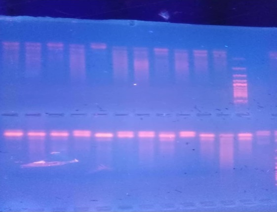
Tissue sections were obtained on a slide, then de-waxed in xylene for two different changes for ten minutes each. They were rehydrated in different grades of alcohols, absolute alcohols (100 percent) for seven minutes, 90% for five minutes, and 70% for three minutes, before being washed in distilled water for two minutes. After re-hydration, slides were stained with hematoxylin for 10 minutes, then blued in running tap water for 15 minutes, counterstained with eosin for 7 minutes, then dehydrated with 70%, 90%, and 100% alcohols. After 2 minutes, each slide was air dried, cleaned in xylene, and mounted with DPX [6]. For DNA Extraction, four micro meter sections of each block was cut and lysed by proteinase K and then precipitated by ethanol or isopropanol. After that, alcohol used to remove unwanted material and cellular debris. Finally, the purified DNA dissolved in water [4]. PCR was performed in a total volume of 25 ul prepared by adding appropriate amounts of reaction components into a 0.5 ml Eppendorf micro tube. The reaction mixture contained 2.5ul of 10x buffer, 1.5 Mm MgCl2, 0.3 Mm of each Dntp, 20 Pm of each general HPV primer and 5Pm of each beta globin primer, two unit of Taq DNA polymerase and 300 ng of template DNA sample extracted from the cases study as positive control. A negative control or blank (all reactants minus target DNA) was used in every run of PCR (Figure 1).
Figure 1. Polymerase Chain Reaction Positive Result of Esophageal Squamous Cells Carcinomain Gel Electrophoresis X40. (Courtesy of Abdelsalam Y Azza and Abdelwadoud Mohamed Elfatih, UMST).

The mixture was overlaid with 40 ul mineral oil and subjected to 40 cycles of PCR amplification using an Eppendorf thermocycler touch down PCR program. At the end pf the PCR, tube was transferred to refrigerator for later use in agarose gel electrophoresis and analysis of the results [4].
The collected data were computerized using an Epi-Info 7 template and analyzed using SPSS 23. The information was summarized quantitatively (mean, standard deviation, and median) as well as graphically (frequency, tables, and graphics). The Fisher test was used to analyze the relationship between categorical variables. All statistical tests were judged significant when the P-value was less than 0.05.
Results and Discussion
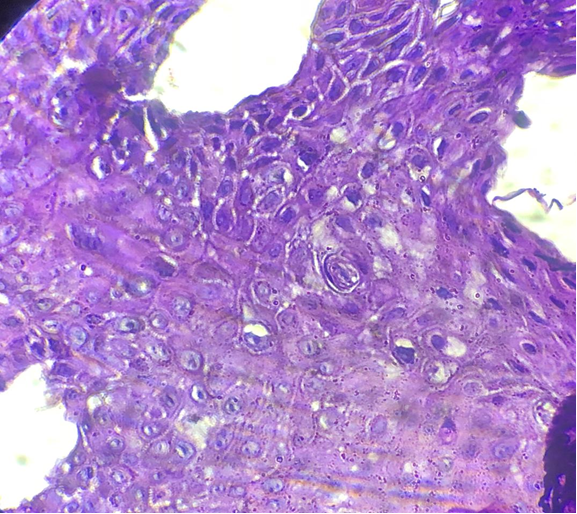
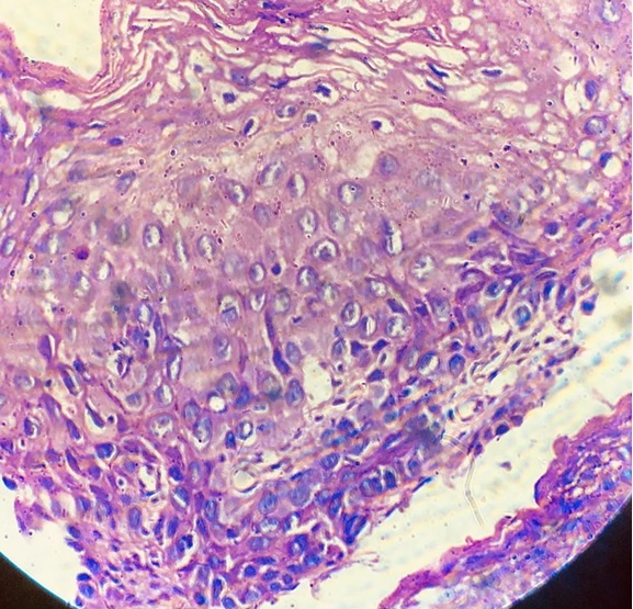
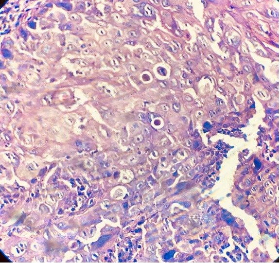
The study assessed the role of HPV-16 in occurrence of ESCCS. The relationship with histological grade, gender and age of esophageal squamous cells carcinoma was investigated in this study. It showed that 38% males (19), 62% females (31), their ages ranged from 17 to 85years with median age of the patients was 70. The percentage of HPV subtypes 16 among the study population was 18%, In relation to the histological grades the detection of HPV 16 was showed in well differentiated squamous cells carcinoma was 11.10% (Figure 2), the moderate differentiated squamous cell carcinoma was 19.20 % (Figure 3) and in the poorly differentiated squamous cell carcinoma was 20.0% (Figure 4) despite the insignificant association between HPV 16 and Esophageal squamous cell carcinoma with P. value 0.
Figure 2. Well Differentiated Squamous Cells Carcinoma of Esophagus H&E X40. (Courtesy of Abdelsalam Y Azza and Abdelwadoud Mohamed Elfatih, UMST).

Figure 3. Moderate Differentiated Squamous Cells Carcinoma of Esophagus with H&E X40. (Courtesy of Abdelsalam Y Azza and Abdelwadoud Mohamed Elfatih, UMST).

Figure 4. Poorly Differentiated Squamous Cells Carcinoma of Esophagus with H&E X40. (Courtesy of Abdelsalam Y Azza and Abdelwadoud Mohamed Elfatih, UMST).

The severe squamous cells carcinoma is predominating the kiolocytic changes and nuclear polymorphism feature of histopathological characteristic in this study. The relationship between human papilloma virus and gender among the study population found that the positive rate of HPV was higher in females (22.6%) than males (10.50%) age ranged from 47 years old to 79 years old.
A total of 50 patients previously diagnosed as esophageal squamous carcinoma 38% males (19), 62% females (31), their ages ranged from 17 to 85 years with median age of the patients was 70 (Table 1).
| Descriptive Statistics | Number | Minimum | Maximum | Median | Mean | Std. Deviation |
| Age (Years) | 50 | 17 | 85 | 70 | 63 | 16 |
| Variables | Number | Percent |
| Age groups | ||
| Less than 40 years | 6 | 12 |
| 40-60 years | 8 | 16 |
| More than 60 years | 36 | 72 |
| Gender | ||
| Male | 19 | 38 |
| Female | 31 | 62 |
| Histological grade | ||
| Well differentiated | 9 | 18 |
| Moderately differentiated | 26 | 52 |
| Poorly differentiated | 15 | 30 |
| HPV16 | ||
| Negative | 41 | 82 |
| Positive | 9 | 18 |
| n=50 |
This finding was consistent with study done in June (2019) A study conducted by AsmaMahir, Mohamed Elbagir, et al who found that the peak of the patients with esophageal cancer was in the age group 65 to 69 years, which constituting 40%. While the positive rate of human papilloma virus high in females’ esophageal cancer was 22.60% [5], Age ranged above 40 and this is not consistent with study [7]. Additional variables of potential prognostic factors such as weight loss, anemia, performance status, dietary habits, sexual behavior, these variables may be the causes of esophageal cancer in this study [8,9].
In this study HPV was detected in 9 (18%) of the cases by using polymerase chain reaction method. This observation was consistent with the previous study [3] [10].
Thecross tabulation between the detection of human papilloma virus subtypes 16 among the study and histological findings showed the positive rate of HPV were in the well differentiated squamous cells carcinoma was 11.10%, while in the moderate differentiated squamous cell carcinoma was 19.20 %, and in the poorly differentiated squamous cell carcinoma was 20.0 %. The histological features of squamous cells carcinoma were characterized by the kiolocytic changes, and nuclear polymorphism.
In conclusion, the positive rate of human papilloma virus subtype-16 were 18%. The most common age of study population observed high positive rate of human papilloma virus were above 40 years old. Females were predominant affected by squamous cells carcinoma. The most common biological differentiation observed high rate of HPV-16 were found in poorly differentiated squamous cells carcinoma. The frequency of HPV-16 was statically insignificant associated by gender, age and histological differentiation.
References
- p63 regulates growth of esophageal squamous carcinoma cells via the Akt signaling pathway S Ye, Kb Lee, Mh Park, Js Lee, Sm Kim. International journal of oncology.2014;44(6). CrossRef
- The aetiological role of human papillomavirus in oesophageal squamous cell carcinoma: a meta-analysis Liyanage Surabhi S., Rahman Bayzidur, Ridda Iman, Newall Anthony T., Tabrizi Sepehr N., Garland Suzanne M., Segelov Eva, Seale Holly, Crowe Philip J., Moa Aye, Macintyre C. Raina. PloS One.2013;8(7). CrossRef
- Human papillomavirus-related esophageal cancer survival Guo Lanwei, Liu Shuzheng, Zhang Shaokai, Chen Qiong, Zhang Meng, Quan Peiliang, Sun Xi-Bin. Medicine.2016;95(46). CrossRef
- Laboratory diagnosis of human papillomavirus virus infection in female genital tract Dixit Rahul, Bhavsar Chintan, Marfatia Y. S.. Indian journal of sexually transmitted diseases and AIDS.2011;32(1). CrossRef
- the prevalence of human papilloma virus in esophageal squamous cell carcinoma Sadat Noori, Ahmed Monabati, Abbasli Ghaderi. Iranian journal of medical sciences.2012;37(2):126-133.
- Detection of human papillomavirus DNA in esophageal carcinoma in Japan by polymerase chain reaction Toh Y., Kuwano H., Tanaka S., Baba K., Matsuda H., Sugimachi K., Mori R.. Cancer.1992;70(9). CrossRef
- Prognostic role of HPV infection in esophageal squamous cell carcinoma Bognár Laura, Hegedűs Ivett, Bellyei Szabolcs, Pozsgai Éva, Zoltán László, Gombos Katalin, Horváth Örs Péter, Vereczkei András, Papp András. Infectious Agents and Cancer.2018;13. CrossRef
- KamangerFarin , Wong Ho and Dawsey Sanford Gastrointestinal clinics of north America,Enivromental causes of esophageal cancer March.10.1016/J.GTC.2009.01.004..
- Gasterointestinal cancer research.Sqaumous cell cancer of the esophagus 2011-Jan-Feb Leichman Lawrence And R.Thomas , JR.CharlesR . ;4(1):22-23.
- HPV infection and p53 and p16 expression in esophageal cancer: are they prognostic factors? Costa Allini Mafra, Fregnani José Humberto Tavares Guerreiro, Pastrez Paula Roberta Aguiar, Mariano Vânia Sammartino, Silva Estela Maria, Neto Cristovam Scapulatempo, Guimarães Denise Peixoto, Villa Luisa Lina, Sichero Laura, Syrjanen Kari Juhani, Longatto-Filho Adhemar. Infectious Agents and Cancer.2017;12. CrossRef
License

This work is licensed under a Creative Commons Attribution-NonCommercial 4.0 International License.
Copyright
© Asian Pacific Journal of Cancer Biology , 2022
Author Details