Bilateral Renal Myelolipoma, Diagnosis Based on Needle Biopsy, Report of a Case
Download
Abstract
Myelolipoma is a rare benign tumor composed of mature adipose tissue and bone marrow-derived hematopoietic elements. The myelolipoma is mostly seen in the adrenal gland. We reported a 53-year old male patient presented with vague abdominal pain. Spiral abdominal and pelvis computed tomography scan showed two masses in both kidneys and most suggestive of lymphoma. Needle biopsy of each mass was taken under guided computed tomography scan and showed mature adipose tissue with hematopoietic elements, especially megakaryocytes-like cells, suggestive of myelolipoma.
Introduction
Myelolipoma is uncommon benign tumor composed of the mature adipose tissue admixed with benign mature hematopoietic elements [1-3]. The most common site of myelolipoma is adrenal gland but may be found in other sites such as pelvis, kidney, retro-peritoneum and thorax [1,4-10].
Extra-adrenal myelolipoma is slightly more common in adult women [1]. The etiologies of extra-adrenal myelolipoma have not yet been determined and there are several theories about embryologic origin and characteristics of chromosomal abnormality with 3q25 translocation to 21p41 and partial deletion of 21 and 17 chromosome short arms [10].
The differential diagnosis of extra-adrenal myelolipoma, depending on the anatomical site, includes retroperitoneal lipoma, liposarcoma and renal angiomyolipoma [5].
The accurate diagnosis of myelolipoma requires histological examination [5,7].
Case report
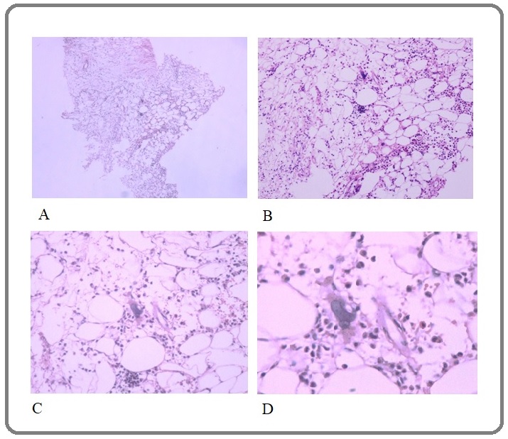
A 53-year old man with insulin-dependent type II diabetes mellitus presented with vague abdominal pain. Physical examination did not reveal any abnormality. Laboratory tests (Blood count, Hemoglobin, Urea and Creatinine) revealed no abnormal findings. Ultrasound examination of the both kidneys showed increased urothelial thickness in the pelvis and ureters more than normal and mild to moderate hydronephrosis. These findings primarily suggested the complications of chronic pyelonephritis, which necessitates further investigations. Computed tomography (CT) of the abdomen and pelvis showed two hypodense masses measuring 110x90 mm in the left kidney and 115x80 mm in the right kidney without enhancement in both kidneys with differential diagnosis of lymphoma and less probably lymphangiectasia and renal sinus lipomatous tumor. The biopsy of the mass under the guided CT was taken. The specimen consisted of multiple needle-shaped fragments measuring totally 0.4x 0.3 cm. Histologically, the specimen composed of mature adipose tissue and hematopoietic elements, small foci of mature lymphoid cells aggregate, RBC and especially megakaryocytes-like cells were noted (Figure1).
Figure 1. Myelolipoma, Hematoxylin-Eosin stain, A) X40, B) X100, C) X200, D) X400 magnifications .

Based on histomorphology, pathology report was “mature adipose tissue with inflammatory cells and megakaryocyte-like elements, compatible with myelolipoma “. For definite diagnosis based on megakaryocyte cells, immunohistochemistry (IHC) including CD34 and CD68 was done but due to lack of these cells in deep section, IHC result was inconclusive. For the definite diagnosis, complete excision was recommended. Written consent was obtained for the report of this case.
Discussion
Myelolipoma is uncommon, benign tumor composed of mature adipose tissue admixed with hematopoietic elements [1-3]. Myelolipoma is mostly found in adrenal gland but may be found in other sites [1]. Myelolipoma doesn’t have specific clinical symptoms and it is often an incidental finding in radiological examination [2,3,11,12]. Internal hemorrhage may be a complication in giant tumors [13]. Some reports of unusual sites of this tumor are discussed in more details in the following.
Qingtong Shi et al., reported primary mediastinal myelolipoma, in a 74-year old male with history of hypertension presented asymptomatic without chest pain, hoarseness, hemoptysis or cough. It was detected as a mediastinal neoplasm on chest X- ray on routine health care. Preoperative diagnosis was difficult but postoperative diagnosis was myelolipoma [10].
Mitual D. Amin et al., reported one case of an unusual site for extra-adrenal myelolipoma in renal sinus of a 66-year old male presented with vague abdominal pain and recurrent urinary tract infections. Differential diagnosis based on radiologic studies included sarcoma (possibly liposarcoma), transitional cell carcinoma and renal cell carcinoma. Fine needle aspiration cytology was interpreted as “consistent with myelolipoma” and finally pathologic examination of nephrectomy specimen confirmed the diagnosis [8].
Kevin S Baker et al., reported a case of presacral myelolipoma. They discussed a case of 79-year old female who presented for evaluation of hip fracture following trauma. On the pelvic computed tomography evaluation showed incidentally heterogenous pre-sacral mass with mixed fat and soft tissue. According to histology, the diagnosis of myelolipoma was made [7].
Min Ho Cho et al., reported another case of presacral myelolipoma in a 70-year old woman presented with persistent lower abdominal pain and anemia. Pelvic magnetic resonance image (MRI) findings were suspicious for liposarcoma but histologic examination of the resected mass suggested pre-sacral myelolipoma [4].
Arsany Hakim and Christoph Rozeik reported thoracic paravertebral myelolipoma . They reported an asymptomatic 70-year old male presented with right paravertebral mass ,had been detected incidentally on chest X- ray. The findings of imaging were presumed diagnosis of extra-adrenal myelolipoma . After guided biopsy, based on histology, diagnosis of myelolipoma was confirmed [5].
Ali Hajiran and the colleagues reported a case of perirenal extra-adrenal myelolipoma in a 78-year old man presented with suspected acute pancreatitis. In the patient work-up a mass was detected in the left retroperitoneal region on computed tomography (CT). Histologic findings after surgical excision suggested perirenal myelolipoma [6].
Merieme Ghaouti and colleagues reported an interesting case similar to the present case. Their case was a male in the sixth decade with insulin-dependent type II diabetes mellitus, renal involvement and hydronephrosis. The difference was the unilaterality in their case [1].
Similar to the above mentioned cases, the present case had no specific symptoms and radiological evaluation showed two masses in the both kidneys. Several radiological differential diagnoses considered such as lymphoma, angiomyolipoma , renal lymphangiectasia and renal sinus lipomatosis. The examination of the biopsy specimen under CT scan was suggestive of myelolipoma.
In conclusion, diagnosis of the extra-renal myelolipoma is difficult because of its less prevalence and lack of the specific clinical symptom. Most of the extra- adrenal myelolipomas are incidentally found on radiologic examination and that have multiple differential diagnoses based on anatomical site. The definite diagnosis of myelolipoma requires histological features after surgical excision or biopsy.
Acknowledgments
The authors would like to thank the Clinical Research Development Center of Imam Reza Hospital for Consulting Services.
References
- Renal myelolipoma: a rare extra-adrenal tumor in a rare site: a case report and review of the literature Ghaouti Merieme, Znati Kaoutar, Jahid Ahmed, Zouaidia Fouad, Bernoussi Zakiya, Mahassini Najat. Journal of Medical Case Reports.2013;7. CrossRef
- Incidental Detection of Adrenal Myelolipoma: A Case Report and Review of Literature Nabi Junaid, Rafiq Danish, Authoy Fatema N., Sofi Ghulam Nabi. Case Reports in Urology.2013;2013. CrossRef
- Adrenal myelolipoma: A rare case report Shanthi Vissa, Rao Nandam M., Chaitanya Balekoduru, Krishna Baddukonda A. R., Mohan Kuppili V. M.. Journal of Dr. NTR University of Health Sciences.2012;1(2). CrossRef
- A case report of symptomatic presacral myelolipoma Cho Min Ho, Mandaliya Rohan, Liang John, Patel Mitesh. Medicine.2018;97(15). CrossRef
- Adrenal and extra-adrenal myelolipomas - a comparative case report Hakim A, Rozeik C. Journal of radiology case reports.2014;8(1). CrossRef
- Perirenal extra-adrenal myelolipoma Hajiran Ali, Morley Chad, Jansen Robert, Kandzari Stanley, Bacaj Patrick, Zaslau Stanley, Cardinal Jon. World Journal of Clinical Cases : WJCC.2014;2(7). CrossRef
- Presacral myelolipoma: a case report and review of imaging findings Baker KS, Lee D, Huang M, Gould ES. Journal of radiology case reports.2012;6(6). CrossRef
- Myelolipoma of the renal sinus. An unusual site for a rare extra- adrenal lesion Amin M. B., Tickoo S. K., Schultz D.. Archives of Pathology & Laboratory Medicine.1999;123(7). CrossRef
- Myelolipoma of the Pelvis: A Case Report and Review of Literature Sethi Seema, Thakur Shivam, Jacques Suzanne, Aoun H. D., Tranchida Paul. Frontiers in Oncology.2018;8. CrossRef
- Primary mediastinal myelolipoma: a case report and literature review Shi Qingtong, Pan Shu, Bao Yang, Fan Huangxin, Diao Yali. Journal of Thoracic Disease.2017;9(3). CrossRef
- A giant myelolipoma discovered as an adrenal incidentaloma: radiological, endocrine and pathological evaluation Sandoval Mark Anthony S., Anel-Quimpo Joselynna. BMJ case reports.2010;2010. CrossRef
- Adrenal myelolipoma: operative indications and outcomes Gershuni Victoria M., Bittner James G., Moley Jeffrey F., Brunt L. Michael. Journal of Laparoendoscopic & Advanced Surgical Techniques. Part A.2014;24(1). CrossRef
- Acute Retroperitoneal Hemorrhage Induced by Giant Adrenal Myelolipoma Mimicking Renal Colic Pain: A Case Report Wu Meng-Yu, Hou Yueh-Tseng, Yiang Giou-Teng. Reports.2018;1(1). CrossRef
License

This work is licensed under a Creative Commons Attribution-NonCommercial 4.0 International License.
Copyright
© Asian Pacific Journal of Cancer Biology , 2022
Author Details