Clinicopathological Evaluation of Gallbladder Lesions in Cholecystectomy Specimens with Special Emphasis on Incidentally Detected Cases of Gallbladder Carcinoma: An Immunohistochemical Study of Cytokeratin 7 and Cytokeratin 20
Download
Abstract
Background and objective: Cholecystectomy specimens exhibit a wide clinicopathological spectrum, ranging from common non-neoplastic diseases to rare neoplastic lesions. Histopathological examination remains the gold standard for definitive diagnosis. Gallbladder cancers (GBCs) are rare, accounting for 0.5% to 1.09% of all gallbladder lesions. These cancers are either clinically suspected or incidentally diagnosed following cholecystectomy.
Materials and Methods: This hospital-based cross-sectional study included all cases diagnosed clinically and radiologically as cholecystitis and subsequently subjected to cholecystectomy. Histopathological diagnosis of gallbladder lesions, age and sex distribution across different gallbladder pathologies, association with gallstones, pathological pT staging of malignant cases, and CK7 and CK20 immunohistochemical findings in malignant cases were observed in this study.
Results: A total of 340 gallbladder specimens were examined histopathologically, revealing 12 cases of gallbladder carcinoma. The most common histopathological diagnosis was chronic cholecystitis (79.4%), followed by chronic cholecystitis with cholesterolosis (7.6%) and adenocarcinoma (3.5%). The most common age group for gallbladder lesions was the fourth decade (26.2%). The age range was 9-85 years, with a mean age of 39.4 years. Overall, females (83%) were more commonly affected than males (17%), with a male:female ratio of 1:4.96. Gallstones were present in 89.1% of all cases, including 83.3% of malignant cases. Incidentally detected gallbladder carcinoma represented 1.17% of the cases, with adenocarcinoma NOS being the most common histopathological type. Pathological pT staging was limited to pT1 and pT2, indicating early-stage disease. CK7 positivity was observed in 91.6% of cases, while CK20 positivity was found in 16.7% of cases. Both CK7 and CK20 were positive in 16.7% of cases, while both were negative in one case (8.33%).
Conclusion: While chronic cholecystitis remains the most common histopathological diagnosis in gallbladder specimens, the possibility of incidental malignancy should be ruled out by mandatory routine histopathological examination of all cholecystectomy specimens.
Introduction
Gallbladder is the organ where bile is stored and concentrated, which helps in digestion of fat [1]. Cholecystectomy is one of the most frequently performed abdominal operation and the gallstones are one of the major causes of morbidity and mortality all over the world affecting 10% of adult population [2]. In India, gallstone disease is seven times more common in the north as compared to the south. Northern and Northeastern states of Uttar Pradesh, Bihar, West Bengal, Orissa, and Assam show the high prevalence of the gall bladder disease. This significant difference was attributed to the environmental factors, diet and lifestyle [3,4].
Cholelithiasis is one of the major gastrointestinal disorders, which is formed due to metabolic problems of hepatobiliary system. It is a major cause of morbidity and mortality throughout the world [5]. The estimated prevalence of gall stone disease in India is 2-29% with 7 times more common in North India than in South India [5,6]. Cholelithiasis cause various changes is gallbladder mucosa ranging from acute cholecystitis, chronic cholecystitis, polyp, empyema, eosinophilic cholecystitis, metaplasia, hyperplasia, dysplasia to carcinoma [7].
Gall Bladder Cancers (GBCs) are rare and account for 0.5% to 1.09% of all gall bladder lesions. It is either clinically suspected or incidentally diagnosed following cholecystectomy for gall stone disease [8].
Cytokeratins are well-described intermediate filament proteins of both normal epithelia and epithelial tumors. Antibodies to several different subtypes of cytokeratin have been available, including cytokeratin 7 (CK7) and cytokeratin 20 (CK20). In normal tissues, CK7 is typically found in simple epithelia from the gastrointestinal tract (including the gallbladder, hepatic ducts and pancreatic ducts), female genital tract (the endometrium and fallopian tube), breast, urinary tract (bladder), and respiratory tract (lung). In contrast, CK20 is found in more complex epithelia from the gastrointestinal tract (such as the gastric and intestinal mucosa), genitourinary tract (urothelium), squamous epithelia from any site, and Merkel cells. Because these cytokeratins usually retain their tissue specificity in their neoplastic counterparts, coordinate expression of these 2 cytokeratins has recently been proposed to help identify the site of origin of various metastatic carcinomas [9].
Studies on various carcinomas suggest that the combined use of Cytokeratin 7 and Cytokeratin 20 may provide helpful information for the discrimination of the origin of metastatic tumours of unknown primary location [10-12]. Again, comparison of the expression pattern of Cytokeratin 7 and Cytokeratin 20 in cases of chronic cholecystitis with lithiasis and in gall bladder carcinoma may highlight possible alterations linking inflammatory and metaplastic changes and carcinoma [13-16].
Aims and Objectives
The aims & objectives of the present study were -
a) To study the histopathological features of different gall bladder lesion with special reference to malignancy.
b) To investigate the co-ordinated expression of Cytokeratin 7 and Cytokeratin 20 in histologically confirmed gall bladder carcinoma.
Materials and Methods
It was a Hospital based cross-sectional Study and included 340 cholecystectomy cases received at the Department of Pathology; Fakhruddin Ali Ahmed Medical College and Hospital during the period of 1 year. Histopathological examination of the gallbladder specimens was done following standard protocols. The specimens those fulfilled the inclusion criteria were included in the study and rest were excluded.
Inclusion criteria
• Formalin fixed cholecystectomy specimens excised for clinically or radiologically diagnosed gall bladder diseases with or without gall stones, polyp or porcelain gallbladder and received in Pathology Department.
Exclusion criteria
• Specimens received in a poorly preserved or autolysed state and improperly labelled were excluded from the study.
The study was conducted after getting permitted by the Institutional Ethical Committee under the IEC no. 10552. All the relevant clinical findings were noted and properly formalin fixed specimens were grossed with three sections each from fundus, body and neck region of the gall bladder. Extra sections were also taken from the tumour or any thickened area of wall in clinically suspicious cases of gallbladder cancer.
Sections were processed and were further subjected to Haematoxylin and Eosin (H&E) stain. H&E stain was done using Harris haematoxyline with a regressive staining method. Sections were examined microscopically and histomorphological evaluation for a wide spectrum of gall bladder lesions was done. The neoplastic lesions were further classified according to the latest 2019 WHO Classification of Digestive System Tumours that includes BilIN, ICPN and Carcinoma [17]. Immunohistochemical examination of malignant cases was done using CK7 & CK20 IHC markers following standard protocols.
Results
Detailed analysis of 340 cholecystectomy specimens was performed under parameters including age, sex, presence of gall stones, histopathological diagnosis, basic pathology of cases, classification of malignant cases with Pathological pT staging, presence of Perineural invasion (PNI) and Lymphovascular invasion (LVI) and CK7 & CK20 immunohistochemical staining status of malignant cases.
A. Histopathological Distribution of the Cases
There were 12 different types of histopathological diagnosis and the most common histopathological diagnosis was chronic cholecystitis (79.4%) followed by chronic cholecystitis with cholesterolosis (7.6%) and adenocarcinoma (3.5%). Incidentally detected gallbladder carcinoma (IGBC) is 1.17%. Details were shown in the Table 1.
| Serial No | Histopathological diagnosis | No of cases | Percentage % |
| 1 | Acute Cholecystitis | 4 | 1.2 |
| 2 | Acute on Chronic Cholecystitis | 2 | 0.6 |
| 3 | Chronic cholecystitis | 270 | 79.4 |
| 4 | Eosinophilic Cholecystitis | 1 | 0.3 |
| 5 | Xanthogranulomatous Cholecystitis | 5 | 1.5 |
| 6 | Chronic Cholecystitis with foreign body giant cell reaction | 3 | 0.9 |
| 7 | Chronic Cholecystitis with epithelial hyperplasia | 4 | 1.2 |
| 8 | Chronic Cholecystitis with adenomatous hyperplasia | 4 | 1.2 |
| 9 | Chronic Cholecystitis with antral metaplasia | 2 | 0.6 |
| 10 | Chronic Cholecystitis with epithelial dysplasia | 7 | 2 |
| 11 | Chronic Cholecystitis with cholesterosis | 26 | 7.6 |
| 12 | Adenocarcinoma | 12 | 3.5 |
| Total | 340 | 100 |
Among the 340 cases, 311 cases (91.4%) were inflammatory lesion, 08 cases (2.3%) cases were hyperplasia, 02 cases (0.6%) were metaplasia, 07 cases (2%) cases were dysplasia and 12 cases (3.5%) were carcinoma.
B. Age Distribution
Age distribution in the present study was from 09 years to 85 years with a mean age of 39.4 years. Again; age distribution in benign lesions of gallbladder cases was from 09 years to 85 years with a mean age of 38.9 years. Overall, the most common age group was 4th decade (26.2%) followed by 3rd decade (24.4%) and 5th decade (23.5%).
C. S ex Distribution
Overall females (83%) were more commonly affected than males (17%) and male: female ratio was 1:4.96. In cholesterosis cases; females (73%) were more commonly affected than male (27%). Again; in both benign and malignant lesions of gallbladder cases; females (83%) were more commonly affected than male (17%). All incidentally detected gallbladder carcinoma (IGBC) cases were observed in females (100%).
D. Association of gallstones in the cholecystectomy cases
Out of total 340 gallbladder specimens; gallstones were found in 303 cases i.e. 89%. Again; out of 303 cases; number of male cases were 52 and females were 251 i.e. 82.8%. Out of 12 malignant cases; gallstones were associated with 10 cases i.e. 83%. Gallstones were present in all 07 gallbladder associated with dysplasia (100%).
E. WHO classification of the malignant cases
There were total 12 number of malignant cases and they were categorized in Table 2.
| WHO Type | ICD-O code | No of cases |
| Adenocarcinoma NOS | 8140/3 | 8 |
| Intracystic papillary neoplasm with associated invasive carcinoma | 8503/3 | 3 |
| Mucinous adenocarcinoma | 8480/3 | 1 |
| Total | 12 |
F. Differentiation of adenocarcinoma NOS cases
There were total 08 number of adenocarcinoma NOS cases and they were classified against their differentiation as:- 06 cases as well differentiated adenocarcinoma and 02 cases as moderately differentiated.
G. Pathological pT staging of the malignant cases
According to their pathological pT staging; the malignant cases were categorized in Table 3.
| Pathological staging | No of malignant cases |
| pT1 | 6 |
| · pT1a | · 01 |
| · pT1b | · 05 |
| pT2 | 5 |
| · pT2a | · 04 |
| · pT2b | · 01 |
| pT3 | 1 |
| pT4 | 0 |
| Total | 12 |
H. Perineural invasion (PNI) & Lymphovascular invasion (LVI) status in malignant case
Out of total 12 malignant cases; PNI was observed in 02 cases and both are diagnosed as moderately differentiated adenocarcinoma and pathological pT staging of the cases were pT2a & pT2b. LVI was not seen in any malignant cases.
I. Incidentally detected gallbladder carcinoma cases (IGBC)
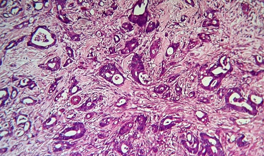
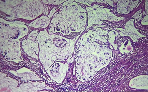
In this study; 04 cases were detected incidentally with incidence rate of 1.17% (04/340) and all cases were female. Age distribution was 25 years to 60 years with a mean age of 46.25 years. The gallbladder wall thickness was ≥ 8 mm in all four cases; which may be one gross finding for gallbladder cancer. Out of total 04 cases; three cases were diagnosed as adenocarcinoma NOS and one case was diagnosed as Intracystic papillary neoplasm with associated invasive carcinoma. Regarding differentiation; 02 cases were diagnosed as well differentiated adenocarcinoma and 01 case as moderately differentiated adenocarcinoma (Figure 1).
Figure 1. Microphotograph of Moderately Differentiated Adenocarcinoma of Gallbladder Showing Malignant Glands Infiltrating the Perimuscular Connective Tissue (40X).
Pathological pT staging of IGBC cases: 02 cases – pT1 and 02 cases – pT2. Details were shown in Table 4.
| Serial No | Age Yrs | Sex | GB wall thickness in millimeter | Histopathological type | Pathological pT staging |
| 1 | 45 | F | 08 mm | Well differentiated adenocarcinoma | pT1b |
| 2 | 55 | F | 08 mm | Intracystic papillary neoplasm with associated invasive carcinoma | pT1a |
| 3 | 25 | F | 15 mm | Moderately differentiated adenocarcinoma | pT2b |
| 4 | 60 | F | 08 mm | Well differentiated adenocarcinoma | pT2a |
J. Immunohistochemistry for CK7 & CK20 findings
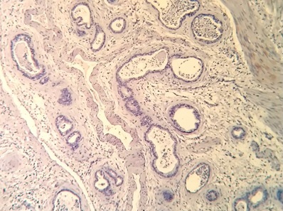
Out of 12 GBC cases; CK7 was positive in 91.6% (11/12) cases and CK20 was positive in 16.7% (2/12) cases. CK20 positive cases were diagnosed as adenocarcinoma NOS with pathological pT staging of pT1. Total number of CK7 & CK20 positive cases were 02, CK7 positive (Figure 2) & CK20 negative (Figure 3) cases were 09 and both negative cases was 01.
Figure 2. Microphotograph of Adenocarcinoma of Gallbladder Showing IHC Marker - CK7 positivity (10X) .
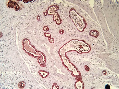
Figure 3. Microphotograph of Above Adenocarcinoma of Gallbladder Showing IHC Marker – CK20 Negative (10X).
Both CK7 & CK20 negative case was diagnosed as mucinous adenocarcinoma (Figure 4 and 5) with pathological staging pT2. Details were shown in Table 5.
Figure 4. Showing Specimen of Gallbladder Having Thickened Gallbladder Wall (case of mucinous adenocarcinoma of gallbladder.
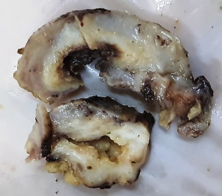
Figure 5. Microphotograph of MucinousAdenocarcinoma of Gallbladder Showing Pools of Extracellular Mucin (40X).
| IHC markers | No of positive cases | Percentage (%) |
| CK7+/CK20+ | 02/12 | 16.70 |
| CK7+/CK20- | 09/12 | 75 |
| CK7-/CK20+ | 00/12 | 0 |
| CK7-/CK20- | 01/12 | 8.33 |
Discussion
In the present study; the most common histopathological diagnosis was chronic cholecystitis (79.4%) which is correlated with other studies like Devi B et al [18] (82%), Mondal B et al [19] (79.8%).
The mean age group of the cholecystectomy cases was 39.4 years which is comparable to Gupta K et al [20] (41.69 years). 4th decade was the commonest age group in the present study; which is similar in the studies by Gupta K et al [20] and Islam Mj et al [21]. The age ranged from 25 years to 65 years in malignant gallbladder cases; which is comparable to Shah B et al [22], Bhattacharjee K P et al [23]. In the present study; the upper limit of age is lower than most of the other studies like Dutta U et al [24], Harikleia K et al [25], Hussain N H et al [26], Geramizadeh B et al [27]etc. The mean age in malignant cases was 49.8 years; which is comparable to other studies like Dutta U et al [24], Hussain N H et al [26], Shah B et al [22], Bhattacharjee K P et al [23].
In the present study; the male:female ratio among the cholecystectomy cases was 1:4.96; which is comparable to the studies - Mondal B et al [19] where male:female ratio was 1:4.2, Kumbhakar D et al [28] (1:4.71), Tiwari A et al [29] (1:4) and Gupta K et al [20] (1:3.9). All incidentally detected gallbladder carcinoma cases were found in females. Gall bladder disease is more common in females attributable to female sex hormones and sedentary lifestyle as the risk factors.
Gall stones cause obstruction that leads to development of chronic cholecystitis which, in turn, chronically predisposes to carcinoma of the gallbladder. Around 83.3% cases of gallbladder carcinoma are associated with gall stones in the present study; which is comparable to Kumar H et al [30] (80%) and Tiwari A et al [29] (80%). The incidence of incidentally detected gallbladder carcinoma was 1.17% which is comparable to the studies like Tiwari A et al [29] (1.25%), Yadav R et al [31] (1.26%). This observation is higher than the other studies like Jetley S et al [32] (0.96%) & Kumbhakar D et al [28] (0.75%).
In the present study, the most common type of carcinoma was adenocarcinoma NOS (66.7%) followed by Intracystic papillary neoplasm with associated invasive carcinoma and this observation is comparable to Giang T H et al [33] (60%), Hussain N H et al [26] (57.6%), Shah B et al [22] (71.4%) and Manuela S et al [34] (65.6%). There were 06 cases of well differentiated adenocarcinoma (75%) followed by 02 cases of moderately differentiated adenocarcinoma and 00 cases of poorly differentiated adenocarcinoma. This observation is comparable to other studies like Dutta U et al [24] (71.4% - Well differentiated adenocarcinoma) and Manuela S et al [34] (52.3% - Well differentiated adenocarcinoma). Perineural invasion (PNI) is noted in 02 cases and no case had shown Lymphovascular invasion (LVI). Both are taken as prognostic parameter for gallbladder carcinoma.
For pathological staging, there were 06 cases in stage pT1 (50%) followed by 05 cases in stage pT2 and one case in stage pT3. This observation is comparable to Siddiqui et al [35] (50% - pT1), Geramizadeh B et al [27] (55.5% - pT1) and Servet K et al [36] (61% - pT1).
Regarding immunohistochemistry findings, out of total 12 cases; 11 cases (91.6%) had shown CK7 positivity; which is higher than other studies like Duval J V. et al [9] (82%), Harikleia K et al [25] (69.05%) and Dursun N et al [37] (57%). Again; 02 cases had shown CK20 positivity (02/12, 16.7%) which is lower than other studies like Duval J V. et al [9] (27%), Harikleia K et al [25] (28.57%) and Dursun N et al [37] (27%).
In conclusion, although the most common gall bladder histopathology diagnosis is chronic cholecystitis, the possibility of any incidental malignancy needs to be ruled out by mandatory routine histopathological examination of all cholecystectomy specimens. Secondly; most of the gallbladder carcinoma cases express CK7 by immunohistochemical examination.
Acknowledgements
This research did not receive any specific grant from funding agencies in the public, commercial, or not-for- profit sectors.
Conflict of Interest
The authors declare no conflict of interest.
References
- Histopathological Study of gall bladder Mehariya MK , Patel MB , Dhotre SV . Int J Res Med.2014;3(4):96-99.
- Histological study of human gallbladder Damor DNT , Chauhan DHM , Jadav DHR . International Journal of Biomedical and Advance Research.2013;4(9). CrossRef
- Original Article - Morphological spectrum of gallstone disease in 1100 cholecystectomies in North India Mohan H, Punia RPS , Dhawan SB , Ahal S, Sekhon MS . 2005.
- Spectrum of clinicopathological presentations of gall bladder diseases in eastern UP Srivastav AC , Srivastava M, Paswan R. International Journal of Contemporary Medicine Surgery and Radiology.2019;4(1):A18-A23.
- Clinical practice. Acute calculous cholecystitis Strasberg SM . The New England Journal of Medicine.2008;358(26). CrossRef
- Cholecystitis Elwood DR . The Surgical Clinics of North America.2008;88(6). CrossRef
- Histopathological spectrum of gall bladder lesions and association with cholelithiasis. International Journal of Medical and Health Research Singh P, et al . 2018;4(9):134-138.
- Cholecystectomies – A 1.5 year histopathological study Kumari NS , Sireesha A, Srujana S, Kumar OS . IAIM.2016;3(9):134-39.
- Expression of cytokeratins 7 and 20 in carcinomas of the extrahepatic biliary tract, pancreas, and gallbladder Duval JV , Savas L, Banner BF . Archives of Pathology & Laboratory Medicine.2000;124(8). CrossRef
- Pathology of the gallbladder, biliary tract, and pancreas. In LiVolsi VA, ed. Major Problems in Pathology Owen DA , Kelly JK . Philadelphia: WB Saunders.2001;39:221-232.
- Pathologic staging of pancreatic, ampullary, biliary, and gallbladder cancers: pitfalls and practical limitations of the current AJCC/UICC TNM staging system and opportunities for improvement Adsay NV , Bagci P, Tajiri T, Oliva I, Ohike N, Balci S, Gonzalez RS , et al . Seminars in Diagnostic Pathology.2012;29(3). CrossRef
- Morphological studies on the gallbladder. II. The “true Luschka ducts” and the “Rokitansky-Aschoff sinuses” of the human gallbladder Halpert B. Bull Johns Hopkins Hosp.1927;41:77-103.
- Cytokeratin 7 and cytokeratin 20 expression in epithelial neoplasms: a survey of 435 cases Chu P, Wu E, Weiss LM . Modern Pathology: An Official Journal of the United States and Canadian Academy of Pathology, Inc.2000;13(9). CrossRef
- Immunohistochemistry as a diagnostic aid in the evaluation of ovarian tumors McCluggage WG , Young RH . Seminars in Diagnostic Pathology.2005;22(1). CrossRef
- CDX2, cytokeratins 7 and 20 immunoreactivity in rectal adenocarcinoma Saad RS , Silverman JF , Khalifa MA , Rowsell C. Applied immunohistochemistry & molecular morphology: AIMM.2009;17(3). CrossRef
- The value of immunohistochemical expression of TTF-1, CK7 and CK20 in the diagnosis of primary and secondary lung carcinomas Al-Zahrani IH . Saudi Medical Journal.2008;29(7).
- Carcinoma of gall bladder. In: Cree IA, editor. WHO classifiation of Tumours of the digestive system 5th ed Rao JC , Adsay NV , Arola J, Tsui WM , Zen Y. Lyon: IARC.2019;:283-88.
- Histopathological Spectrum of Diseases in Gallbladder Devi B , et al . National Journal of Laboratory Medicine.2017;6(4):PO06-PO09.
- Histopathological spectrum of gallstone disease from cholecystectomy specimen in rural areas of West Bengal, India- an approach of association between gallstone disease and gallbladder carcinoma Mondal B, Maulik D, Biswas BK , Sarkar GN , Ghosh D. International Journal Of Community Medicine And Public Health.2016;3(11). CrossRef
- The spectrum of histopathological lesions in Gallbladder in cholecystectomy specimens Gupta K, Faiz A, Thakral R, Mohan A, Sharma V. International Journal of Clinical and Diagnostic Pathology.2019;2. CrossRef
- Role of Routine Histopathology of Gallbladder Specimen from Gallstone Disease to Detect Unsuspected Carcinoma Islam M, Akter S, Talukder A, Haque M. Journal of Bangladesh College of Physicians and Surgeons.2019;37. CrossRef
- A retrospective audit of gall bladder histopathology following cholecystectomy Shah B, Degloorkar S. IP Journal of Diagnostic Pathology and Oncology.2020;3(2).
- Prospective observational study on cholelithiasis in patients with carcinoma gall bladder in a tertiary referral hospital of Eastern India Bhattacharjee PK , Nanda D. Journal of Cancer Research and Therapeutics.2019;15(1). CrossRef
- Clinicopathological evaluation of gallbladder carcinoma with special emphasis on incidentally detected cases- A hospital based study Dutta U, Saikia P, Gogoi G, Borgohain M. Indian Journal of Pathology and Oncology.2019;6. CrossRef
- Cytokeratin 7 and 20 expression in gallbladder carcinoma Kalekou H, Miliaras D. Polish Journal of Pathology: Official Journal of the Polish Society of Pathologists.2011;62(1).
- Clinicopathological study of gall bladder carcinoma with special reference to gallstones: our 8-year experience from eastern India Hamdani NH , Qadri SK , Aggarwalla R, Bhartia VK , Chaudhuri S, Debakshi S, Baig SJ , Pal NK . Asian Pacific journal of cancer prevention: APJCP.2012;13(11). CrossRef
- Incidental Gall Bladder Adenocarcinoma in Cholecystectomy Specimens; A Single Center Experience and Review of the Literature Geramizadeh B, Kashkooe A. Middle East Journal of Digestive Diseases.2018;10(4). CrossRef
- A Histopathological Study of Cholecystectomy Specimens Kumbhakar D. Journal of Medical Science And clinical Research.2016. CrossRef
- Importance of Routine Histopathological Examination of Gallbladder Specimen in Detecting Incidental Malignancies Tiwari A, Rai R, Jain SK . J Lumbini Med Coll.2016;4(1):15-19.
- Histological evaluation of 400 cholecystectomy specimens Kumar H, Kini H, Tiwari A. Journal of Pathology of Nepal.2015;5:834-840.
- Incidental Gallbladder Carcinoma in North Indian Population: Importance of Routine Histopathological Examination of All Benign Gallbladder Specimens Yadav R, Sagar M, Kumar S, Maurya SK . Cureus.2021;13(7). CrossRef
- Incidental gall bladder carcinoma in laparoscopic cholecystectomy: a report of 6 cases and a review of the literature Sujata J, S R, Sabina K, Mj H, Jairajpuri ZS . Journal of clinical and diagnostic research: JCDR.2013;7(1). CrossRef
- Carcinoma involving the gallbladder: a retrospective review of 23 cases - pitfalls in diagnosis of gallbladder carcinoma Giang TH , Ngoc TT , Hassell LA . Diagnostic Pathology.2012;7. CrossRef
- Hyperplasia, metaplasia, dysplasia and neoplasia lesions in chronic cholecystitis - a morphologic study Stancu M, Căruntu ID , Giuşcă S, Dobrescu G. Romanian Journal of Morphology and Embryology = Revue Roumaine De Morphologie Et Embryologie.2007;48(4).
- Routine histopathology of gallbladder after elective cholecystectomy for gallstones: waste of resources or a justified act? Siddiqui FG , Memon AA , Abro AH , Sasoli NA , Ahmad L. BMC surgery.2013;13. CrossRef
- Preneoplastic and neoplastic gallbladder lesions detected after cholecystectomy Kocaöz S, Turan G. Przeglad Gastroenterologiczny.2019;14(3). CrossRef
- Mucinous carcinomas of the gallbladder: clinicopathologic analysis of 15 cases identified in 606 carcinomas Dursun Nevra, Escalona Oscar Tapia, Roa Juan Carlos, Basturk Olca, Bagci Pelin, Cakir Asli, Cheng Jeanette, Sarmiento Juan, Losada Hector, Kong So Yeon, Ducato Leslie, Goodman Michael, Adsay N. Volkan. Archives of Pathology & Laboratory Medicine.2012;136(11). CrossRef
License

This work is licensed under a Creative Commons Attribution-NonCommercial 4.0 International License.
Copyright
© Asian Pacific Journal of Cancer Care , 2023
Author Details
How to Cite
- Abstract viewed - 0 times
- PDF (FULL TEXT) downloaded - 0 times
- XML downloaded - 0 times