Epithelioid Hemangioma of the Spine: A Rare Case Report and Review of Literature
Download
Abstract
Background and objective: Epithelioid hemangioma (EH) of the spine is a rare vascular disease. Hemangiomas are tumors typically composed of thin-walled blood vessels, with EH representing an uncommon variant. While considered benign, EH can be locally aggressive. Although most commonly affecting the integumentary system, EH can also involve the liver, lungs, and bones. This case report presents a 25-year-old male with a comprehensive workup for EH, including clinical data, MRI spine, surgical findings, histopathological information, and adjuvant radiation therapy following surgical management. In addition to the case report, a review of the literature on EH of bones is provided.
Case Presentation: A 25-year-old male presented to our emergency department with complaints of severe lower back pain and difficulty walking for the past three months. A previous MRI of the lumbosacral (LS) spine with whole spine screening, performed one month prior, revealed multifocal vertebral lesions involving the D4, D5, and D6 vertebrae, predominantly in the posterior elements. Expansion of the transverse processes, involvement of the costochondral junction, and the left 5th rib with extensive soft tissue component indenting on the pleura were also observed. The patient was evaluated by the neurosurgery team, and surgical decompression and stabilization of the D4-D6 vertebrae were planned. He underwent a D3-D6 laminectomy and stabilization under general anesthesia. Final histopathology, along with immunohistochemistry (IHC) correlation, confirmed a diagnosis of epithelioid hemangioma. The case was discussed at our hospital’s multidisciplinary tumor board, and adjuvant radiotherapy (RT) was recommended. The patient received a total dose (TD) of 45 Gray (Gy) to the D3-D6 region. Six months post-treatment, his power has improved and is near normal.
Conclusion: This case exhibits typical features of EH, including a young male patient, lytic lesions with a sclerotic rim, and IHC confirmation.
Introduction
Epitheloid hemangioma (EH) of spine is a rare vascular disease. Haemangiomas are tumours typically composed of thin walled blood vessels, of which EH is an infrequent variation [1]. The exact incidence of EH is not known. EH, though is considered to be benign disease it can be locally aggressive. EH most commonly affects the integumentary system. IT most commonly involves liver, lungs and bones. Several other sites have been described in the literature like lymph nodes, lung, breasts. The term was first descried by Dail and Liebow in the year 1975. According to the recent WHO classification of bone tumours, EH is defined as low to intermediate grade malignant neoplasm. Bone is the second most commonly affected site [2,3]. It can involve long tubular bones of extremity, phalanges of hands and foot, axial skeleton. Sometimes, EH can be misdiagnosed as multiple myeloma, metastatic lesion from some other sites etc. Diagnosis is usually made with histopathology and IHC correlation [4-6]. Treatment of EH includes surgery, radiation or surveillance [7,8]. Radiation therapy is mostly used in the adjuvant setting (After surgery) [9,10]. There is not much published literature on EH due to the rarity of the disease. Here we report a 25 years old male with complete work up for EH including clinical data, MRI spine, surgical, histopathological information and adjuvant radiation therapy following surgical management. In addition to the case report, we reviewed literature on EH of bones. The aim of this study is to guide clinicians in diagnosing EH with clinical information, imaging features and in treatment.
Case Report
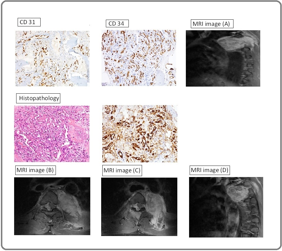
A 25 years old male came to our emergency department with complaints of severe lower back pain. He also complained of difficulty in walking for the past 3 months. No history of fever, cough, loss of weight, loss of appetite, bowel/bladder disturbances. He was evaluated outside for the same. MRI Lumbosacral (LS) with whole spine screening was done 1 month back and it showed Multifocal vertebral lesions involving D4, D5, D6 vertebra predominantly in posterior elements, expansion of the transverse processes and involvement of costochondral junction, left 5th rib with extensive soft tissue component indenting on the pleura. He was referred to our hospital for further evaluation and management. On examination, performance status 3, B/L lower power limb power 1/5. MRI LS spine was repeated as his condition had worsened and it showed T1 isointense, T2 heterointense lobulated expansile lesion noted involving D4 right hemi body, transverse process, left 5th rib adjacent costo chondral junction with soft tissue component indenting pleura and D6 right hemi body, pedicle, tranverse process. Epidural components were noted at D4, D5, D6 levels causing significant canal narrowing compressing the spinal cord at D6 level (MRI images A, B, C, D) (Figure 1).
Figure 1. Showing MRI Images and Histopathological Images along with IHC.

He was seen by Neurosurgery team at our hospital and was planned for surgical decompression and stabilisation of D4-D6 vertebrae. He underwent D3 to D6 laminectomy and stabilization under general anaesthesia. Procedure was uneventful. Histopathology was reported as vascular neoplasm and further IHC for keratin, CD31, CD 34, ERG, p53 and Ki67 were requested. All IHC except p53 showed positive reaction. Ki67 was 10 % (Figure 1). Final histopathology along with IHC correlation was suggestive of Epitheloid Hemangioma. Case was discussed in our hospital multidisciplinary tumor board and was planned for adjuvant radiotherapy (RT). Then he received adjuvant radiotherapy total dose (TD) 45 Gray (Gy) to D3-D6 region. He was simulated in supine position with vacloc for immobilisation. He was treated with conformal 3D technique. He was treated with 3 field technique. The target volume received 97 percent of prescribed dose. He was treated with 1.8 Gy per fraction, five days a week to a total dose of 45Gy in 25 fractions. His lower limb power improved during the course of radiotherapy to 3/5. Tolerated treatment well. No radiotherapy induced reactions. Post treatment he is on regular follow up. Post treatment 6 months, his power is improved and is near normal.
Discussion and Review of Literature
EH is a rare vascular tumour arising from endothelial or pre endothelial cells. The most common presenting symptom is pain. The neurological symptoms correlate with the location of mass. The age at presentation is between 20 to 30 years, the median age being 25 years. It has a male predominance with a male to female ratio being 2:1. A study done by Kerry and his colleagues reported an equal incidence of EH among males and females. Rosai et al proposed a disease model consisting of previously described diseases of skin, soft tissues, bone and heart [11,12]. EH can be metastatic as well [13]. The histologic similarity of EH to angiolymphoid hyperplasia with esonopihilia (ALHE) suggests that both are representative of single neoplastic entity which was later termed as histiocytic hemangioma [14]. EH most commonly occurs in metaphysis and diaphysis of long tubular bones of extremities and followed by short tubular bones of extremities. Patients usually present with back pain and neurological symptoms. It can also occur in forehead, pre auricular area, scalp and lacrimal gland [15], inner canthus of eye [16], heart [17], penis [18], scrotum [19], testis [20], colon [21], lymph nodes [22],
breasts [23], tongue [24] and spleen [14]. Among cases of EH arising in bone only 16% occurred in vertebra even in the largest series of 50 cases of EH reported till date. Multi-modality imaging plays a critical role in assessment and management of patients with EH. On X ray or CT scan, the typical characteristics are osteolytic lesion with well-defined margin. EH of bone is usually defined by an MRI. MRI also shows a lytic lesion with a sclerotic rim. The signal intensity is not specific, such as low to intermediate signal intensities in T1 weighted images; High signal intensity on T2 weighted images and restricted diffusion. A halo sign can be found after administration of contrast. Our patient had lesions in multiple spine. Similar imaging manifestations like multifocal presentation, aggressive radiologic appearance may be present in both benign and malignant conditions. It has to be distinguished from epithelioid hemangioendothelioma (EHE) and angiosarcoma. Therefore, imaging should not be considered definitive for diagnosing EH. Identifying microscopic features along with IHC correlation is of utmost importance in diagnosing EH. The morphologic features depend on the formation stage, location as well as presence of vascular and inflammatory components. IHC plays an important role in diagnosing EH. The important IHC markers include CD 31, CD 34 FLI-1, ERG, FOSB, CAMTA-1, TFE-3, INI-1 and keratin. Multifocal lesions of bone EH are frequently seen. The clear distinction between multifocal EH and metastatic disease does not exist. Treatment options include combinations of biopsy, surgical resection, pre operative embolization and adjuvant radiotherapy. There is very less information regarding role of RT in EH. To the best of our knowledge, only 2 cases of EH of vertebra so far have received RT till date. Our patient was diagnosed with EH of spine and underwent surgical resection followed by adjuvant RT. Kerry et al and Albakr et al suggested that most common treatment for EHE of spine was surgical resection followed by adjuvant RT [25]. Luzzati et al reported that patients who had undergone wide resection had a better prognosis [26]. Adjuvant RT is recommended to reduce the local recurrence after surgery. Our patient was pain free and neurologically improved during follow up period of 6 months after multimodality treatment.
In conclusion, to summarize our case appears as a typical features of EH like young male patient, lytic lesions with sclerotic rim, IHC confirmation. However imaging features are not specific of EH. MRI remains gold standard for identifying EH of spine. Follow up MRI scans are necessary to rule out new lesions in spine.
References
- Epithelioid hemangioma of bone and soft tissue: a reappraisal of a controversial entity Errani C, Zhang L, Panicek DM , Healey JH , Antonescu CR . Clinical Orthopaedics and Related Research.2012;470(5). CrossRef
- WHO Classification of Tumours of Soft Tissue and Bone. 4th ed. Lyon: IARC; 2013 Fletcher CDM , Bridge JA , Hogendoorn PCW , Mertens FE , editors . .
- Epithelioid hemangioma of the spine: Two cases O'Shea BM , Kim J. Radiology Case Reports.2014;9(4). CrossRef
- Epithelioid hemangioma of bone. A tumor often mistaken for low-grade angiosarcoma or malignant hemangioendothelioma O'Connell JX , Kattapuram SV , Mankin HJ , Bhan AK , Rosenberg AE . The American Journal of Surgical Pathology.1993;17(6).
- ZFP36-FOSB fusion defines a subset of epithelioid hemangioma with atypical features Antonescu CR , Chen H, Zhang L, Sung Y, Panicek D, Agaram NP , Dickson BC , Krausz T, Fletcher CD . Genes, Chromosomes & Cancer.2014;53(11). CrossRef
- Frequent FOS Gene Rearrangements in Epithelioid Hemangioma: A Molecular Study of 58 Cases With Morphologic Reappraisal Huang S, Zhang L, Sung Y, Chen C, Krausz T, Dickson BC Brendan C., Kao Y, et al . The American Journal of Surgical Pathology.2015;39(10). CrossRef
- Epithelioid hemangioma of the spine: a case series of six patients and review of the literature Boyaci B, Hornicek FJ , Nielsen GP , DeLaney TF , Pedlow FX , Mansfield FL , Carrier CS , Harms J, Schwab JH . The Spine Journal: Official Journal of the North American Spine Society.2013;13(12). CrossRef
- Vascular tumors of bone: A study of 17 cases other than ordinary hemangioma, with an evaluation of the relationship of hemangioendothelioma of bone to epithelioid hemangioma, epithelioid hemangioendothelioma, and high-grade angiosarcoma Evans HL , Raymond AK , Ayala AG . Human Pathology.2003;34(7). CrossRef
- Radiotherapy for large symptomatic hemangiomas Schild SE , Buskirk SJ , Frick LM , Cupps RE . International Journal of Radiation Oncology, Biology, Physics.1991;21(3). CrossRef
- Epithelioid hemangioma of bone revisited: a study of 50 cases Nielsen GP , Srivastava A, Kattapuram S, Deshpande V, O'Connell JX , Mangham CD , Rosenberg AE . The American Journal of Surgical Pathology.2009;33(2). CrossRef
- The histiocytoid hemangiomas. A unifying concept embracing several previously described entities of skin, soft tissue, large vessels, bone, and heart Rosai J, Gold J, Landy R. Human Pathology.1979;10(6). CrossRef
- Angiolymphoid hyperplasia with eosinophilia. A clinicopathologic study of 116 patients Olsen TG , Helwig EB . Journal of the American Academy of Dermatology.1985;12(5 Pt 1). CrossRef
- Epithelioid hemangioma of bone: a potentially metastasizing tumor? Floris G, Deraedt K, Samson I, Brys P, Sciot R. International Journal of Surgical Pathology.2006;14(1). CrossRef
- Epithelioid haemangioma (angiolymphoid hyperplasia with eosinophilia) in the inner canthus Mariatos G, Gorgoulis VG , Laskaris G, Kittas C. Journal of the European Academy of Dermatology and Venereology: JEADV.2001;15(1). CrossRef
- Lacrimal gland epithelioid haemangioma Coombes AG , Manners RM , Ellison DW , Evans BT . The British Journal of Ophthalmology.1997;81(11). CrossRef
- Solid epithelioid hemangioma of the heart: Report of a case with unusual threadlike bridging strands. Koren J , Zamecnik M . Journal of Interdisciplinary Histopathology.2015;3(4):135-137.
- Epithelioid hemangioma of the penis: case report and review of literature Ismail M, Damato S, Freeman A, Nigam R. Journal of Medical Case Reports.2011;5. CrossRef
- Epithelioid hemangioma-a rare scrotal tumor of childhood Avallone AN , Avallone MA , Share S, Rubin BP . Urology.2012;80(3). CrossRef
- Epithelioid hemangioma of the testis Liu X, Wang R, Guan W, Wang L. Indian Journal of Pathology & Microbiology.2013;56(4). CrossRef
- Epithelioid hemangioma of the colon: a case report Nonose R, Priolli DG , Cardinalli IA , Máximo FR , Galvão PSP , Martinez CAR . Sao Paulo Medical Journal = Revista Paulista De Medicina.2008;126(5). CrossRef
- Primary nodal hemangioma Elgoweini M, Chetty R. Archives of Pathology & Laboratory Medicine.2012;136(1). CrossRef
- Vascular proliferations of the breast Brodie C, Provenzano E. Histopathology.2008;52(1). CrossRef
- Epithelioid hemangioma of the tongue mimicking a malignancy Shimoyama T, Horie N, Ide F. Journal of Oral and Maxillofacial Surgery: Official Journal of the American Association of Oral and Maxillofacial Surgeons.2000;58(11). CrossRef
- Hemangioendothelioma arising from the spleen: A case report and literature review Wang Z, Zhang L, Zhang B, Mu D, Cui K, Li S. Oncology Letters.2015;9(1). CrossRef
- Epithelioid hemangioendothelioma of the spine: case report and review of the literature Albakr A, Schell M, Drew B, Cenic A. Journal of Spine Surgery (Hong Kong).2017;3(2). CrossRef
- Epithelioid hemangioendothelioma of the spine: results at seven years of average follow-up in a series of 10 cases surgically treated and a review of literature Luzzati A, Gagliano F, Perrucchini G, Scotto G, Zoccali C. European Spine Journal: Official Publication of the European Spine Society, the European Spinal Deformity Society, and the European Section of the Cervical Spine Research Society.2015;24(10). CrossRef
License

This work is licensed under a Creative Commons Attribution-NonCommercial 4.0 International License.
Copyright
© Asian Pacific Journal of Cancer Care , 2023
Author Details
How to Cite
- Abstract viewed - 0 times
- PDF (FULL TEXT) downloaded - 0 times
- XML downloaded - 0 times