Prognostic Significance of Double-Expresser Status in Diffuse Large B-cell Lymphoma: Insights from a Tertiary Care Cancer Centre in India
Download
Abstract
Background: Diffuse large B-cell lymphoma (DLBCL) is characterized by the proliferation of medium to large B lymphoid cells with a diffuse histopathologic growth pattern. The presence of the double-expresser (DE) phenotype, defined by co-expression of MYC and BCL2 proteins via immunohistochemistry (IHC), has been associated with inferior survival in DLBCL. This study aimed to assess the prevalence of DE status in DLBCL and evaluate its prognostic value.
Methods: A retrospective analysis was conducted in a tertiary care cancer centre, focusing on all DLBCL, NOS cases diagnosed in 2012. MYC and BCL2 protein expression was determined using IHC. The prognostic significance of double-expressers was evaluated by comparing the progression-free survival (PFS) and overall survival (OS) between double-expressers and non-double expressers, employing appropriate statistical methods. Data were analyzed using SPSS version 11, and survival probabilities were estimated using the Kaplan-Meier method. The log-rank test was employed to assess differences in survival among various prognostic factors. Prognostic factors were further evaluated using univariate and multivariate Cox-regression models. A P-value < 0.05 was considered statistically significant.
Results: DE lymphoma accounted for 22.2% (n=18) of all DLBCL, NOS cases. DE status was associated with significantly shorter PFS (P-value = 0.049) and OS (P-value = 0.015).
Conclusion: The presence of DE status is indicative of poor prognosis in DLBCL, NOS. Assessment of MYC and BCL2 protein expression via IHC provides a rapid and cost-effective approach to risk-stratify DLBCL patients at the time of diagnosis.
Introduction
Diffuse large B-cell lymphoma (DLBCL) is the most common type of non-Hodgkin lymphoma (NHL). They are heterogeneous group of tumours with diverse clinical and biological behaviour and are subdivided into morphological variants, molecular subtypes, and distinct disease entities based on morphology, cell of origin, immunophenotype, and genetic profile [1, 2].
DLBCL with MYC and BCL-2 protein co-expression by immunohistochemistry are categorized as double-expresser lymphomas (DEL). The co-expression of MYC and BCL2 proteins should be considered a prognostic biomarker of poor clinical outcome. Co- expression of these proteins is predictive of poor prognosis at diagnosis and relapse. DEL comes under DLBCL not otherwise specified (DLBCL-NOS) category in the 2017 revised 4th edition WHO classification of Tumours of haematopoietic and lymphoid tissues. In DEL, two-thirds of patients belong to the activated B- cell (ABC) subtype and one-third belong to germinal centre B- cell (GCB) subtype [1, 3]. The prognostic and predictive factors described in DLBCL include specific clinical features, morphology, immunophenotype, proliferation index, genetic factors, tumour microenvironment, microRNA expression patterns host genetics, and treatment regimens explaining the variable clinical outcome [3-6]. Objectives of this study were to assess the double-expression status in DLBCL and to assess the utility of double-expression status in predicting the prognosis in patients of DLBCL by measuring the progression free survival (PFS) and overall survival (OS).
Materials and Methods
This was a retrospective study conducted in a tertiary care cancer centre. Study population included all the cases of DLBCL, NOS diagnosed in 2012. Study was approved by IRB and ethical committee (HEC No. 19/2019). Inclusion criteria for the study was defined as cases of DLBCL, NOS diagnosed in the cancer centre in 2012. Exclusion criteria was defined as cases of DLBCL, NOS, slides and blocks of which could not be retrieved from the archives, cases with inadequate tissue and cases diagnosed in the cancer centre by histopathology but complete lymphoma workup, staging and treatment were done in another centre. The sample size was estimated based on the study by Riedell PA et al and the estimated minimum sample required for the present study was 81 [7, 8]. Of the 184 cases diagnosed in 2012 and included according to the inclusion criteria, we excluded 44 cases as per the exclusion criteria. From the remaining cases, 81 were selected for this study by computer-generated random sampling. The slides and blocks of selected cases were retrieved and reviewed. MYC and BCL2 expressions were analysed by immunohistochemistry (IHC). The details of cases selected for the study were collected using a proforma. Details collected include registration number, biopsy number, age, sex, Ann Arbor stage, date of diagnosis, LDH score, ECOG performance status, extra nodal disease status, bone marrow involvement, progression status, recurrence status, follow- up dates and the date of death if the patient had expired. The prognostic significance of double-expressers was analysed by comparing progression- free survival (PFS) and overall survival (OS) of double-expressers and non-double expressers using appropriate statistical methods. PFS was taken from the date of diagnosis to the date of event (progression/ recurrence /death) or date of last follow- up. OS was taken from the date of diagnosis to the date of death or date of last follow- up. In order to reduce the number of lost to follow-up cases, we attempted to contact the patients through phone, and data were collected. IHC was done on additional tissue sections taken from the retrieved blocks. Two separate sections were taken for c-MYC and BCL2. Anti-bcl-2 (SP66 clone) Rabbit Monoclonal Primary Antibody is directed against human bcl-2 expressed by B-cells of the mantle zone and interfollicular T-cells. This antibody exhibits a cytoplasmic staining pattern. The c-MYC (EP121) Rabbit Monoclonal Primary Antibody is directed against oncogene-encoded protein c-MYC. This antibody exhibits a nuclear staining pattern. Both the antibodies are intended for qualitative staining in sections of formalin-fixed, paraffin-embedded tissues. IHC staining was done by automation in VENTANA Bench Mark XT. BCL2 expression was considered positive when ≥50% of tumour cells were positive and c-MYC was considered positive when ≥40% of tumour cells were positive [1]. Data collection was done by retrieving the case sheets of the 81 cases. The details of the cases selected for the study were entered into the proforma for analysis. The follow-up details were accessed from case records or by directly enquiring via phone. All data were analysed using SPSS 11 software. Continuous variables were represented by the mean and standard deviation. Categorical variables were expressed using frequency and relative proportion. The associations between two categorical variables were assessed using Chi-square test/ Fisher’s Exact Test. Kaplan-Meier method was used to estimate the survival probability. A significant difference in survival between various prognostic factors was tested using the log-rank test. Prognostic factors were assessed using univariate and multivariate Cox-regression model. A P- value < 0.05 was considered to be statistically significant. Measures were also taken to eliminate bias. Selection bias was avoided by selecting cases by computer generated random numbers. The pathologist was made unaware of the outcome while assessing the double-expresser status to exclude observer bias.
Results
In the study group, 22.2% (n = 18) were DEL. Comparison of clinical, laboratory parameters and survival of DEL and Non-DEL is shown in Table 1.
| Non-DEL | DEL | P-value | |
| Age (> 60years) n (%) | 18 (28.60 %) | 8 (44.40 %) | 0.255 |
| Male female ratio | 1.52 | 1.25 | 0.789 |
| CNS involvement | 4 (6.30 %) | nil | 0.57 |
| Bone marrow involvement n (%) | 69 (9.50 %) | 3 (16.70 %) | 0.408 |
| LDH mean | 1031.2 | 1667.4 | 0.099 |
| Advanced Stage n (%) | 41 (65.10 %) | 16 (88.90 %) | 0.051 |
| High-intermediate and High IPI score n (%) | 17 (27.00 %) | 10 (55.60 %) | 0.045 |
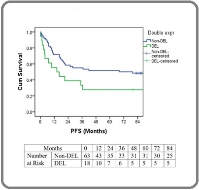
| Progression-free Survival Probability 5 years (%) | 51.8% (SE= 6.5%) | 27.8% (SE= 10.6%) | 0.049 |
| Progression-free Survival Probability 7 years (%) | 48.4% (SE=6.5%) | 27.80% (SE=10.6%) | 0.049 |
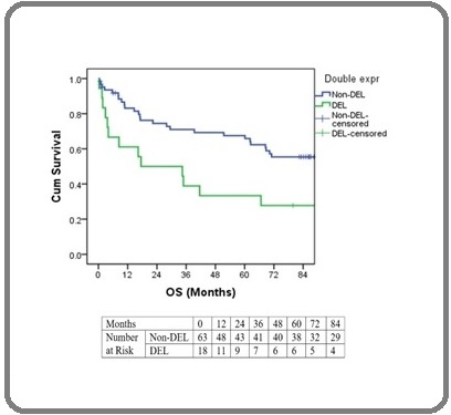
| Overall survival probability 5 years (%) | 65.8% (SE=6.2%) | 33.3% (SE=11.1%) | 0.015 |
| Overall survival probability 7 years (%) | 55.4% (SE=6.5%) | 27.8% (SE=10.6%) | 0.015 |
Age of the patients in the study group ranged from 15 to 86 yrs. Median age of the DEL was 58 years. In the study, male to female ratio was 1.45:1. Males were predominantly affected in both DEL and Non-DEL. Advanced stage disease was present in 88.8% of DEL and 65.1% of Non- DEL. CNS involvement was seen in 6.3% (n=4) of non-DEL and was absent in DEL. Bone Marrow involvement was seen in 9.5% (n=6) of non-DEL and 16.7% (n=3) of DEL. Most common morphological variant in both DEL and Non-DEL group was centroblastic variant. The mean LDH value was higher in DE than in non-DE, but was not statistically significant. In this study, DE predominantly presented with high – intermediate and high IPI score (n=10, 55.6%) whereas non-DEL predominantly presented with low and low- intermediate score (n=46, 73%) and this difference was found to be statistically significant (P-value = 0.045). Median follow-up period was 92 months. The 5- year PFS was 46.2% (SE=5.6%) and 7- year PFS was 43.7% (SE= 5.6%). PFS was less in DEL group when compared to Non-DEL and was found to be statistically significant (P- value 0.049) (Figure 1).
Figure 1. Comparison of Progression Free Survival in DEL and Non-DEL .

The 5- year OS was 58.2% (SE=5.6%) and 7- year OS was 49% (SE=5.7%). Overall survival was less in DEL group and was found to be statistically significant (P-value=0.015) (Figure 2).
Figure 2. Comparison of Overall Survival in DEL and Non-DEL .

Univariate analysis indicated that stage is a significant independent prognostic factor for PFS (Table 2).
| Variables | P-value | HR | 95.0% CI for HR | |
| Lower | Upper | |||
| Age (>60 vs ≤ 60) | 0.051 | 1.831 | 0.996 | 3.365 |
| Sex (Female vs Male) | 0.97 | 1.012 | 0.557 | 1.838 |
| Double-expresser (DEL vs Non-DEL) | 0.053 | 1.896 | 0.991 | 3.629 |
| Stage (III & IV vs I & II) | 0 | 5.336 | 2.094 | 13.597 |
| Bone marrow (Involved vs Not Involved) | 0.952 | 0.972 | 0.383 | 2.467 |
| CNS (Involved vs Not Involved) | 0.258 | 1.815 | 0.646 | 5.098 |
| Morphology (Centro blast vs Immuno blast) | 0.507 | 0.617 | 0.148 | 2.566 |
| Morphology (anaplastic vs Immunoblast) | 0.992 | 1.012 | 0.091 | 11.207 |
| IPI (Poor vs good) | 0.136 | 1.588 | 0.865 | 2.917 |
Age and DE status were found to be marginally significant. Univariate analysis done for OS indicated that age, stage, IPI and DE status were significant independent prognostic factors (Table 3).
| P- value | Hazard rate (HR) | 95% CI for HR | ||
| Lower | Upper | |||
| Age (>60 vs ≤ 60) | 0.015 | 2.171 | 1.164 | 4.052 |
| Sex (Female vs Male) | 0.77 | 1.096 | 0.593 | 2.027 |
| Double-expresser (DEL vs Non- DEL) | 0.018 | 2.226 | 1.149 | 4.311 |
| Stage (III & IV vs I & II) | 0.001 | 4.857 | 1.898 | 12.43 |
| Bone marrow (Involved vs Not Involved) | 0.875 | 1.078 | 0.423 | 2.748 |
| CNS (Involved vs Not Involved) | 0.21 | 1.939 | 0.688 | 5.464 |
| Morphology (Centro blast vs Immuno blast) | 0.373 | 0.523 | 0.125 | 2.181 |
| IPI (Poor vs good) | 0.045 | 1.891 | 1.014 | 3.527 |
Multivariate Cox regression was done with significant covariates obtained in univariate analysis, however only stage turned out to be significant and hence multivariate analysis does not exist.
Discussion
In the present study, the frequency and prognostic significance of DE status in DLBCL, NOS were analyzed. The frequency of DEL in DLBCL, NOS in this study was 22.2% (n=18). The difference in frequency values in published literature may possibly be due to different cut-off values for c-MYC and BCL-2 positivity, different antibody clones and difference in study population selection. The frequency of DEL in our study is almost similar to that mentioned in most other studies [9-12]. Kawashima et al showed a much greater frequency as compared to others because their population was composed exclusively of patients with de novo DLBCL or DLBCL transformed from follicular lymphoma, who underwent allogenic haematopoietic stem cell transplantation [13]. In this study median age of study group was 53 years, and the median age among double-expressers was 58years. The median age of DEL in the study is almost similar to that mentioned in other studies [13, 14]. In this study majority of the cases were males as seen in other similar studies. There is a slight male predominance in both DLBCL, NOS and DEL. In this study, 55.60% of DEL had high-intermediate-/high risk IPI score, whereas only 27.7% of Non-DEL belonged to high-intermediate/high-risk group. This was found to be statistically significant. From the results of this study as well as the other studies, it can be seen that most of the DEL cases have a high-intermediate/high IPI score [9, 15, 16]. In this study, mean LDH at diagnosis for all patients was higher in DEL as compared to Non-DEL but was not found to be statistically significant. Similar results can be seen in other studies also [9, 15]. DEL characteristically presents in advanced stage at the time of diagnosis [9, 10, 12]. In this study, out of the 18 cases of DEL, 88.9% of the cases presented at stage III or IV. The P- value was found to be 0.051. In this study, 88.9% of DEL had morphology of centroblastic variant, and 11.1% had morphology of immunoblastic variant. In a similar study, 60% had centroblastic variant morphology and 25% had immunoblastic variant morphology among DEL [9]. Centroblastic variant shows a predominance in both the studies. Immunoblastic variant was also predominantly seen in DEL in both these studies. In the study, 16.7% of the DEL showed bone marrow involvement as compared to 9.5% in Non- DEL, and was not statistically significant. The frequency of bone marrow involvement was similar to that mentioned in other studies [10, 12, 17]. In this study, the 5-year PFS for DEL was found to be 27.8% (SE= 10.6%) as compared to 51.8% (SE= 6.5%) for Non-DEL. Also, the 7-year PFS for DEL was found to be 27.8% (SE=10.6%) as compared to 48.4% (SE=6.5%) for Non-DEL. This was found to be statistically significant. Comparison of PFS in various studies revealed that PFS for DEL ranged from 20-32% [10, 12, 13]. In the above studies, PFS was shorter for DEL when compared to Non-DEL and was found to be statistically significant. In this study, the 5-year OS for DEL was found to be 33.3% (SE=11.1%) as compared to 65.8% (SE=6.2%)
for Non-DEL. Also, the 7-year OS for DEL was found to be 27.8% (SE=10.6%) as compared to 55.4% (SE=6.5%) for Non-DEL. This difference in OS was found to be statistically significant. Comparison of OS in various studies showed that OS for DEL ranged from 22-47% [9, 10, 12, 13, 16]. In the above studies, DEL showed inferior OS when compared to Non-DEL and was found to be statistically significant. Limitation of this study was that FISH analysis for MYC, BCL2 and BCL6 rearrangements to exclude DHL and THL cases were not done.
To conclude, the frequency of DEL in DLBCL, NOS is 22.2%. DEL presented more often in advanced stage and showed higher LDH values and high- intermediate/ high risk IPI score. Difference in IPI score between DEL and Non- DEL was statistically significant. DEL showed significantly inferior OS and PFS when compared to Non- DEL. Double-expresser status is associated with poor prognosis in DLBCL, NOS. Assessment of MYC and BCL2 expression by IHC represents a rapid and inexpensive approach to risk-stratify patients with DLBCL at the time of diagnosis.
References
- eds. WHO Classification of Tumours of Haematopoietic and Lymphoid Tissues. Revised 4th ed. Lyon, France: International Agency for Research on Cancer Swerdlow SH , Campo E, Harris NL , Jaffe ES , Pileri SA , Stein H, et al . 2017;:p. 291-297.
- A case of a diffuse large B-cell lymphoma of plasmablastic type associated with the t(2;5)(p23;q35) chromosome translocation Adam P, Katzenberger T, Seeberger H, Gattenlöhner S, Wolf J, Steinlein C, Schmid M, et al . The American Journal of Surgical Pathology.2003;27(11). CrossRef
- Approach to the diagnosis and treatment of high-grade B-cell lymphomas with MYC and BCL2 and/or BCL6 rearrangements Sesques P, Johnson NA . Blood.2017;129(3). CrossRef
- Double-hit and double-protein-expression lymphomas: aggressive and refractory lymphomas Sarkozy C, Traverse-Glehen A, Coiffier B. The Lancet. Oncology.2015;16(15). CrossRef
- Quick reference handbook for surgical pathologists. Berlin Heidelberg: springer; 2011 Aug 27:46-51. Rekhtman N, Bishop JA . .
- Low absolute lymphocyte count is a poor prognostic factor in diffuse-large-B-cell-lymphoma Cox MC , Nofroni I, Ruco L, Amodeo R, Ferrari A, La Verde G, Cardelli P, et al . Leukemia & Lymphoma.2008;49(9). CrossRef
- Double hit and double expressors in lymphoma: Definition and treatment Riedell PA , Smith SM . Cancer.2018;124(24). CrossRef
- Practical issues in calculating the sample size for prevalence studies Naing L, Winn T, Rusli BN . Archives of orofacial Sciences.2006;1:9-14.
- Double Hit and Double Expresser Diffuse Large B Cell Lymphoma Subtypes: Discrete Subtypes and Major Predictors of Overall Survival Mehta A, Verma A, Gupta G, Tripathi R, Sharma A. Indian Journal of Hematology & Blood Transfusion: An Official Journal of Indian Society of Hematology and Blood Transfusion.2020;36(4). CrossRef
- MYC/BCL2 protein coexpression contributes to the inferior survival of activated B-cell subtype of diffuse large B-cell lymphoma and demonstrates high-risk gene expression signatures: a report from The International DLBCL Rituximab-CHOP Consortium Program Hu S, Xu-Monette ZY , Tzankov A, Green T, Wu L, Balasubramanyam A, Liu W, et al . Blood.2013;121(20). CrossRef
- The Frequency of Double Expresser in Selected Cases of High Grade Diffuse Large B-Cell Lymphomas Naseem M, Asif M, Khadim MT , Ud-Din H, Jamal S, Shoaib I. Asian Pacific journal of cancer prevention: APJCP.2020;21(4). CrossRef
- Concurrent expression of MYC and BCL2 in diffuse large B-cell lymphoma treated with rituximab plus cyclophosphamide, doxorubicin, vincristine, and prednisone Johnson NA , Slack GW , Savage KJ , Connors JM , Ben-Neriah S, Rogic S, Scott DW , et al . Journal of Clinical Oncology: Official Journal of the American Society of Clinical Oncology.2012;30(28). CrossRef
- Double-Expressor Lymphoma Is Associated with Poor Outcomes after Allogeneic Hematopoietic Cell Transplantation Kawashima I, Inamoto Y, Maeshima AM , Nomoto J, Tajima K, Honda T, Shichijo T, et al . Biology of Blood and Marrow Transplantation: Journal of the American Society for Blood and Marrow Transplantation.2018;24(2). CrossRef
- Prognostic impact of diffuse large B-cell lymphoma with extra copies of MYC, BCL2 and/or BCL6: comparison with double/triple hit lymphoma and double expressor lymphoma Huang S, Nong L, Wang W, Liang L, Zheng Y, Liu J, Li D, Li X, Zhang B, Li T. Diagnostic Pathology.2019;14(1). CrossRef
- Outcome of patients with double-expressor lymphomas (DELs) treated with R-CHOP or R-EPOCH. Blood. 2016;128(22):5396. Aggarwal A, Rafei H, Alakeel F, Finianos AN , Liu ML , El-Bahesh E, et al . .
- Double or triple-expressor lymphomas: prognostic impact of immunohistochemistry in patients with diffuse large B-cell lymphoma Peña C, Villegas P, Cabrera ME . Hematology, Transfusion and Cell Therapy.2020;42(2). CrossRef
- Immunohistochemical double-hit score is a strong predictor of outcome in patients with diffuse large B-cell lymphoma treated with rituximab plus cyclophosphamide, doxorubicin, vincristine, and prednisone Green TM , Young KH , Visco C, Xu-Monette ZY , Orazi A, Go RS , Nielsen O, Gadeberg OV , Mourits-Andersen T, Frederiksen M, Pedersen LM , Møller MB . Journal of Clinical Oncology: Official Journal of the American Society of Clinical Oncology.2012;30(28). CrossRef
License

This work is licensed under a Creative Commons Attribution-NonCommercial 4.0 International License.
Copyright
© Asian Pacific Journal of Cancer Care , 2024
Author Details
How to Cite
- Abstract viewed - 0 times
- PDF (FULL TEXT) downloaded - 0 times
- XML downloaded - 0 times