Beyond the Usual Suspects: A Case Series and Review of Rare Lung Carcinomas
Download
Abstract
Lung cancer remains a significant public health concern, with non-small cell lung carcinoma (NSCLC) and small cell lung carcinoma (SCLC) being the most prevalent subtypes. However, a spectrum of rare lung carcinomas exists, posing unique diagnostic and therapeutic challenges. These rare entities often mimic the clinical presentation of more common subtypes, making early suspicion and timely diagnosis crucial for optimal patient management. This manuscript presented a case series of three rare lung carcinomas, highlighting the importance of recognizing and diagnosing these less frequent malignancies. The clinical features, diagnostic workup, treatment approaches, and prognostic implications specific to each case were also discussed. Moreover, this review provided a comprehensive overview of the current literature on rare lung carcinomas, emphasizing their distinctive characteristics and emphasizing the need for a high index of suspicion among clinicians. This case series underscores the importance of a multidisciplinary approach involving histopathological analysis, immunohistochemistry, and advanced imaging studies in the diagnosis and management of rare lung cancers. Early recognition and appropriate treatment strategies are critical for improving patient outcomes and addressing the complexities of this heterogeneous group of malignancies.
Introduction
Lung cancer includes a variety of tumours depending upon the cell of origin. There are several types of lung carcinoma which are often referred to as subtypes of lung carcinoma. Few of them are found commonly and some are very rare. The two key subtypes of lung cancer include Non-small cell carcinoma (NSCLC) and Small cell lung carcinoma (SCLC). NSCLC is the most common variety. According to the American Cancer Society (ACS), NSCLC accounts for 80-85% of lung carcinomas. SCLC is responsible for approximately 10-15% of cases [1]. The treatment approaches for these two types of cancers differ from each other. Apart from these entities, there are several types of lung cancers which are rare varieties and are encountered infrequently in clinical practice. Examples of such tumours include Adenosquamous carcinoma of the lung, large cell neuroendocrine tumour, salivary gland type lung carcinoma, Lung carcinoids, and Mesotheliomas. In adults, mediastinal tumors can include Germ cell tumors, lymphomas, thymomas, etc. Other extremely rare forms of lung cancer include sarcomatoid carcinoma of the lung and malignant granular cell lung tumors [2]. The symptoms or clinical presentation of such rare tumors may be similar to the commoner varieties.
However, their diagnosis is a clinical challenge in common practice. Treatment and outlook of these tumors also differ from common varieties. In this case series we describe a series of such 3 rare subtypes of lung cancers diagnosed in our clinical practice at a tertiary care centre.
Case 1
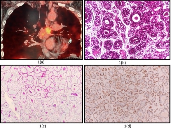
A 63 year old male, non-smoker presented to our department with complaints of cough, breathlessness, and streaky hemoptysis of 03 months duration. On evaluation, his vital parameters were within normal range. His chest auscultation revealed absent breath sounds in the left hemithorax. His routine haematological and biochemical parameters were normal. The chest radiograph showed left sided pleural effusion. A computed tomography (CT) chest was performed which showed soft tissue mass in the left main bronchus (LMB) measuring 38x24x31 mm with left sided pleural effusion. He underwent PET CT which revealed an endobronchial mass in LMB measuring 2.5X3.9X3.2 cm with SUV max of 6.53 (Figure 1a) with a pleural effusion on left side along with FDG avid mediastinal lymph nodes at left hila with SUV of 5.63 and subcarinal station with SUV of 7.30 .
Figure 1. (a) Endobronchial Mass in LMB Measuring 2.5X3.9X3.2 cm with SUV Max of 6.53 (b) H and E stain (400x magnification). Tumor Arranged in varying sized tubules lined by inner ductal and outer basal cells.(c) PAS stain with diastase highlights the magenta colored basement membrane around the individual tubules as well as within the tubule (d) IHC with smooth muscle actin (cytoplasmic) highlights the outer basal (myoepithelial cells).

He was taken up for bronchoscopy and a large growth completely occluding the left main bronchus (LMB) was seen. An endobronchial biopsy was performed which revealed adenoidcystic carcinoma positive for CK7, EMA and CD117 (Figure 1b,c,d). He was started on 4 cycles of chemotherapy (injection paclitaxel, injection carboplatin) and concurrent radiotherapy with 60 Gy/30 #. He is doing well and is on treatment for the past 6 months.
Case 2
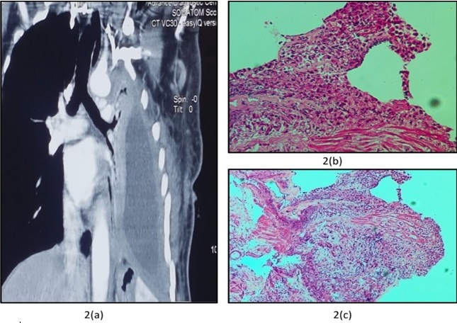
A 65 year old female, non -smoker became symptomatic with productive cough, breathlessness, and weight loss of 3 months duration. On examination, she had tachycardia (pulse rate of 108/min) and tachypnea (respiratory rate of 26/min). On chest examination breath sounds were absent in the left hemithorax. Her hematological and biochemical parameters were normal. CXR showed pleural effusion of the left side and CT chest showed pleural effusion with collapse of the left lung (Figure 2a).
Figure 2. (a) CT Chest Showing Pleural Effusion (left) with Underlying Collapsed Lung. (b and C), Bronchoscope guided biopsy from secondary carina showing adenosquamous carcinoma lung.

She underwent intercostal tube drainage. Pleural fluid was exudative, lymphocyte predominant with ADA of 30 IU/L. A medical thoracoscopy and biopsy revealed fibrinous pleuritis. She was started on anti- tubercular therapy. However, the Patient showed poor response to treatment and after 2 months was referred to our centre. She underwent bronchoscopy which showed congested secondary carina left side with extrinsic compression. Her biopsy from the secondary carina revealed adenosquamous carcinoma (Figure 2b,c) with CK7 and P63 positive. Her PET CT revealed metastasis to the spine and liver. She was started on cisplatin- based chemotherapy.
Case 3
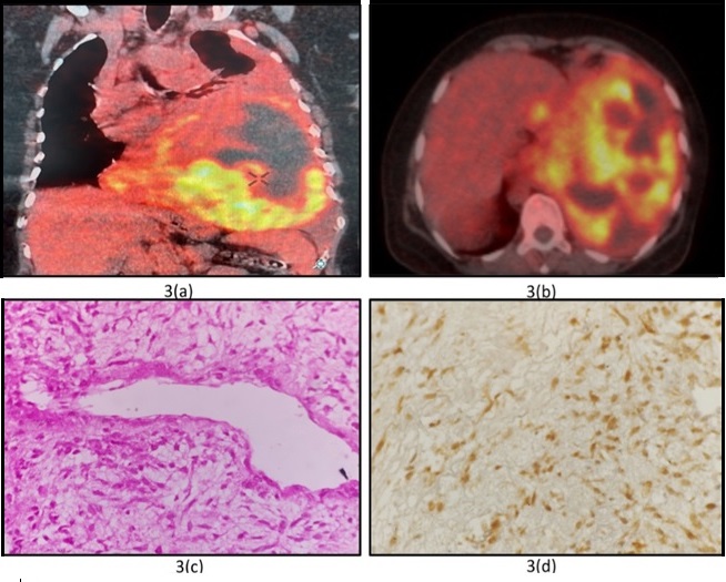
A 54 year old female, non-smoker with no known co-morbidities presented with complaints of cough, and breathlessness of 3 weeks duration. On clinical evaluation, she had tachypnea (respiratory rate-34/min), tachycardia (pulse rate-120/min) and saturation of 94% at ambient air. Her chest auscultation revealed absent breath sounds of left hemithorax. Her chest radiograph revealed massive pleural effusion left side with the tracheal shift to the right. A diagnostic pleurocentesis was performed which revealed exudative haemorrhagic effusion with ADA of 5 U/L. Her pleural fluid malignant cytology and cell block were negative. She underwent a CT chest which revealed pleural effusion and collapsed lung, however no lung mass was discernible. A PET CT was performed which showed a large heterogeneously FDG avid solid (30-50 Hounsfield Unit) and cystic (16-30HU) density mass lesion noted which was involving the entire left hemithorax (Figure 3a).
Figure 3. (a) large heterogeneously FDG avid solid (30-50 HU) and cystic (16-30HU) density mass lesion noted involving almost the entire left hemithorax. (b). The lesion measured 16.8AP x 16.7TR x 15.8CC cm (SUV max-10.78). (c) H and E Stain (400x magnification) show the tumor cells to have moderately pleomorphic elongated wavy hyperchromatic nuclei with accentuation around delicate capillaries. (d) S100p IHC (400X magnification) shows the tumor cells to be positive (nucleocytoplasmic).

The mass was causing collapse consolidation of the entire left lung with relative sparing of the left upper lobe. This lesion was causing a mass effect resulting in contralateral mediastinal shift. Midline crossing was noted at the level of DV10. The lesion measured 16.8AP x 16.7TR x 15.8CC cm (SUV max-10.78) (Figure 3b,c). FDG avid pleural effusion was noted in the upper 1/3rd of the left hemithorax (SUV max-3.98). The lesion was abutting the chest wall with no obvious infiltration. Inferiorly the lesion involved the left hemidiaphragm causing a caudal shift and downward displacement of the spleen. A CT- guided biopsy was done which showed sarcoma (Figure 3c, d).The Patient was started on platinum based chemotherapy and radiotherapy. She is currently on treatment for 4 months.
Discussion
Lung cancer remains a leading cause of mortality and morbidity related to cancers. The commonest histological variety was squamous cell carcinoma, however in recent times a rise in adenocarcinoma and small cell carcinoma has been seen. In developed countries, adenocarcinoma has already replaced squamous cell carcinoma as the predominant histological variant. Also, in a study in north India, an increase in adeno carcinoma has been reported with a prevalence of both squamous and adeno being equal (36.4%) [3]. Apart from common subtypes of lung cancer, there are clinical entities that are very rare in occurrence and differ in diagnostic workup and treatment. The definition of rare cancer varies in different regions. In Europe and Asia, a malignancy has been considered rare if it affects <6 out of 100,000 people yearly. However, the National Cancer Institute of the United States considers cancer rare if it occurs in <15 out of 100,000 people annually [4].
We described three such cases of rare lung cancers that had similar clinical presentation as the common varieties of lung carcinomas. The three cases presented above were adenoid cystic carcinoma, sarcoma and adenosquamous carcinoma.
Case 01 in our series was diagnosed to have Adenoid cystic carcinoma (ACC), a malignant neoplasm originating from salivary glands. It commonly originates from the gland of the head & neck region however incidences of origin from the skin, breast, lungs, and upper digestive tract are also reported in the literature. Earlier this lung malignancy was referred to as benign glandular neoplasm, however now it is classified as a low- grade bronchial carcinoma. Primary lung ACC (ACCL) is a rare form accounting for 0.04-0.2% of all primary lung tumours [5]. Hence due to its rarity, Primary Lung ACC has been reported only in case studies or small case series. Hu MM et al presented retrospective data of 34 cases of ACCL collected over a stretch of over 20 years for this rare variety. They found that ACCL tends to occur in non-smoker, young patients, with an approximate male/female ratio of 1:1 [6]. Clinical presentations are usually cough, hemoptysis, and breathlessness and they are commonly treated as asthma or bronchitis till the time radiological workup is done. The site of origin of ACCL is the trachea-bronchial glands in the submucosa of airways, with a similar morphology of ACC arising elsewhere in salivary glands. ACCL exhibits three different histological growth patterns. The most frequent pattern is the cribriform followed by tubular and least frequent however most aggressive form is of solid pattern. On immunohistochemistry, the expression of myoepithelial markers, such as P63, SMA, C-kit, and S-100 protein, is a strong argument for ACC of the lung [7]. The prognosis of ACCL depends on the tumor stage, histological subtype, and surgical margin status. Among the treatment options of ACCL surgical resection remains the optimal management. For residual disease control and recurrence prevention radiotherapy is indicated for unresectable tumors and incomplete resection [8]. It may show partial response to targeted therapy and the role of Imatinib has been shown in strongly c-Kit expressing tumors [9].
Adenosquamous carcinoma of the lung (ASC) is another distinct histological subtype that is rare and poorly described. ASC is a rare subtype of NSCLC, and it is defined as a malignancy comprising components of lung adenocarcinoma (ADC) and lung squamous cell carcinoma (SCC). Case 02 in our series was diagnosed with adenosquamous carcinoma. ASC accounts for 0.4%– 4% of all lung carcinomas [10]. Naunheim K S et al in their study found 2.3 % out of 873 patients had adenosquamous carcinoma [11]. Maeda et al identified 114 ASC cases in their prospective analysis of 4,668 postoperative patients with pulmonary malignancy. The ratio of male to female according to their paper was 3.38:1 [12]. High rates (74.4%–88.5%) of smoking in patients with ASC have been confirmed by many studies. According to the proportional components on histology, ASC is further subdivided into 03 subtypes: Adenocarcinoma (ADC) - predominant ASC (the proportion of ADC ≥60% of tumor), Squamous cell carcinoma (SCC)-predominant ASC (the proportion of SCC ≥60% of tumor) and structure-balanced ASC (the proportion of ADC and SCC is between 40% and 60%). Surgery is the preferred treatment option for ASC patients. Platinum-based doublet chemotherapy should be considered as a standard treatment option depending on the staging of NSCLC. EGFR-TKIs can be used as a treatment modality for ASC found to be EGFR-positive. In cases of advanced ALK-rearrangement, Crizotinib may be a potential choice [13]. Takamori et al found poorer outcomes in post-surgical cases of ASC as compared to adenocarcinoma and squamous cell carcinoma [14].
Primary sarcomas of the lung (PSL) constitute a heterogeneous group of rare malignant tumors, accounting for less than 0.5% of malignant lung tumors. They arise from mesenchymal tissue of the bronchial wall vessels or pulmonary stroma and are usually present in middle-aged to elderly individuals [15]. The most common subtypes on a histological basis are synovial sarcoma, leiomyosarcoma, epitheloid hemangioendothelioma, and malignant peripheral nerve sheath tumor. They are followed by undifferentiated pleomorphic sarcoma, liposarcoma, and rhabdomyosarcoma. Lung metastasis from extrapulmonary sarcomas is more common than primary pulmonary sarcomas [16]. Although primary lung sarcoma is a diagnosis of exclusion immunohistochemistry may help with keratin, EMA, CD34, actin, and desmin found positive. Primary therapeutic modality includes wide surgical resection aiming for tumor-free margins. Neoadjuvant chemotherapy based on anthracycline and ifosfamide has been disappointing in primary lung tumors. Chemoradiation may be the best therapeutic strategy in unresectable lesions. Collaud S et al in their single centre retrospective study achieved 5 years survival of 60% among their 13 PPS patients treated with induction before curative-intent surgery as part of multimodality management [17].
In conclusion, since these cancers are rare a high index of suspicion and prior knowledge of these varieties may aid in successful and early diagnosis. Due to the paucity of large clinical trials on treatment and management of such cases personalized precision therapy may be warranted in such cases for favourable outcomes. An early PET CT may be useful in discerning the lesion. There is also a felt need for a clinical registry of such rare carcinoma cases in Asian countries.
References
- What Is Lung Cancer? | Types of Lung Cancer [Internet]. [cited 2024 Feb 26]. Available from: https://www.cancer.org/cancer/types/lung-cancer/about/what-is.html .
- Rare lung cancers Breathe.2015;11(4). CrossRef
- Evolving epidemiology of lung cancer in India: Reducing non-small cell lung cancer-not otherwise specified and quantifying tobacco smoke exposure are the key Kaur H, Sehgal IS , Bal A, Gupta N, Behera D, Das A, Singh N. Indian Journal of Cancer.2017;54(1). CrossRef
- Rare molecular subtypes of lung cancer Harada G, Yang S, Cocco E, Drilon A. Nature Reviews. Clinical Oncology.2023;20(4). CrossRef
- Pulmonary adenoid cystic carcinoma: molecular characteristics and literature review Chen Z, Jiang J, Fan Y, Lu H. Diagnostic Pathology.2023;18(1). CrossRef
- Primary adenoid cystic carcinoma of the lung: Clinicopathological features, treatment and results Hu M, Hu Y, He J, Li B. Oncology Letters.2015;9(3). CrossRef
- Histopathologic and Cytogenetic Features of Pulmonary Adenoid Cystic Carcinoma Roden AC , Greipp PT , Knutson DL , Kloft-Nelson SM , Jenkins SM , Marks RS , Aubry MC , García JJ . Journal of Thoracic Oncology: Official Publication of the International Association for the Study of Lung Cancer.2015;10(11). CrossRef
- Adenoid cystic carcinoma of the airway: thirty-two-year experience Maziak DE , Todd TR , Keshavjee SH , Winton TL , Van Nostrand P, Pearson FG . The Journal of Thoracic and Cardiovascular Surgery.1996;112(6). CrossRef
- Primary adenoid cystic carcinoma of lung: a case report and review of the literature Bhattacharyya T, Bahl A, Kapoor R, Bal A, Das A, Sharma SC . Journal of Cancer Research and Therapeutics.2013;9(2). CrossRef
- Clinical characteristics and prognosis of patients with lung adenosquamous carcinoma after surgical resection: results from two institutes Zhu L, Jiang L, Yang J, Gu W, He J. Journal of Thoracic Disease.2018;10(4). CrossRef
- Adenosquamous lung carcinoma: clinical characteristics, treatment, and prognosis Naunheim KS , Taylor JR , Skosey C, Hoffman PC , Ferguson MK , Golomb HM , Little AG . The Annals of Thoracic Surgery.1987;44(5). CrossRef
- Adenosquamous carcinoma of the lung: surgical results as compared with squamous cell and adenocarcinoma cases Maeda H, Matsumura A, Kawabata T, Suito T, Kawashima O, Watanabe T, Okabayashi K, Kubota I. European Journal of Cardio-Thoracic Surgery: Official Journal of the European Association for Cardio-Thoracic Surgery.2012;41(2). CrossRef
- Adenosquamous carcinoma of the lung Li C, Lu H. OncoTargets and Therapy.2018;11. CrossRef
- Clinicopathologic characteristics of adenosquamous carcinoma of the lung Takamori S, Noguchi M, Morinaga S, Goya T, Tsugane S, Kakegawa T, Shimosato Y. Cancer.1991;67(3). CrossRef
- Primary Sarcoma of the Lung - Prognostic Value of Clinicopathological Characteristics of 26 Cases Duran-Moreno J, Kokkali S, Ramfidis V, Salomidou M, Digklia A, Koumarianou A, Tomos P, Koufopoulos N, Vamvakaris I, Psychogiou E, Syrigos K. Anticancer Research.2020;40(3). CrossRef
- Primary sarcomas of the lung: a clinicopathological and immunohistochemical study of 14 cases Attanoos RL , Appleton MA , Gibbs AR . Histopathology.1996;29(1). CrossRef
- Multimodality treatment including surgery for primary pulmonary sarcoma: Size does matter Collaud S, Stork T, Schildhaus H, Pöttgen C, Plönes T, Valdivia D, Zaatar M, Dirksen U, Bauer S, Aigner C. Journal of Surgical Oncology.2020;122(3). CrossRef
License

This work is licensed under a Creative Commons Attribution-NonCommercial 4.0 International License.
Copyright
© Asian Pacific Journal of Cancer Care , 2024
Author Details
How to Cite
- Abstract viewed - 0 times
- PDF (FULL TEXT) downloaded - 0 times
- XML downloaded - 0 times