A Rare Case of Bilateral Ovarian Adult Granulosa Cell Tumors Associated with Uterine Lipoleiomyoma- Case Report and Review of Literature
Download
Abstract
Leiomyomas are the most common uterine tumors and uterine lipoleiomyoma is a rare variant. Lipomatous tumors of the uterus includes spectrum of lesions comprising of lipomas, lipoleiomyoma, angiomyolipomas and fibrolipomyomas in the benign category and liposarcomas in the malignant category. Sex cord-stromal tumors are uncommon ovarian neoplasms comprising of both benign and malignant entities with a wide spectrum of clinical presentation owing to hormonal manifestations. We present a case of a post menopausal female who complained of bleeding and had an incidental bilateral ovarian tumor, which turned out to be adult granulosa cell tumor of the ovary.
Introduction
Leiomyomas are the most common uterine tumors which have a varied presentation [1]. However, uterine lipoleiomyoma are quite rare compared to other variants [2]. The incedence of lipoleoimyomas is about 0.03-0.2% [3]. Lipomatous tumors of the uterus are rare, includes spectrum of lesions comprising of lipomas, lipoleiomyoma, angiomyolipomas and fibrolipomyomas in the benign category and liposarcomas in the malignant category [4].
Leiomyomas are seen to be associated with varied incidentally occuring concurrent lesions such as granulosa cell tumor of ovary (3.65%), dermoid cyst (0.49%), mucinous (4.14%), serous cystadenoma of ovary (5.99%), chocolate cyst of ovary (0.8%), and infective lesions such as tubercular endometritis and salpingitis [5].
Sex cord-stromal tumors are uncommon ovarian neoplasms comprising of both benign and malignant entities, comprising 5% to 12% of ovarian tumors [6]. They can be divided into two subtypes, namely adult and juvenile types based on histological findings and are known to produce estrogen The incidence of adult granulose cell tumor is 2-5% of all ovarian malignancy [7]. Sex cord stromal tumors have a wide spectrum of clinical presentation owing to hormonal manifestations. All tumors must thus be microscopically evaluated to rule out the mimics [8].
Case Report
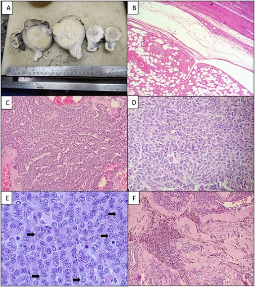
Fifty three year old post-menopausal lady presented bleeding. Per vaginal examination revealed bulky uterus with bilateral adnexal mass and no involvement of the parametrium. CT scan done in an outside centre showed a hyperechoic intramural mass suggestive of leiomyoma measuring approximately 5.8X5X4 cm along with bilateral solid adnexal masses. That there were no metastatic lymph nodes. Total abdominal hysterectomy with bilateral salpingo-oophorectomy was performed. Grossly, the uterus was increased in size, which on cut section showed a well circumscribed mass measuring 6.7x5.5x4.2 cm and the cut section was solid, predominantly yellowish intermixed with whitish whorled areas. Left and right ovarian mass measured 4.5x3.7x 3.5 cm and 3.6x3.2x2.5 cm respectively (Figure 1A).
Figure 1. A) Gross Picture Showing Bilateral Solid Ovarian Tumors Along with Uterine Lipoleiomyoma B) Lipoleiomyoma composed of mature adipocytes admixed with fascicles of smooth muscle (H and E,100X) C) Adult granulosa cell tumor showing tumor cells arranged in trabecular pattern (H and E, 100X) D) Call Exner bodies are evident E) Many of the cells show characteristic nuclear grooving (arrow) F) Focal areas show stromal sclerosis and hemosiderin laden macrophages.

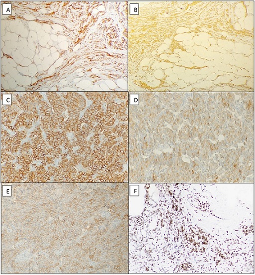
Surface was smooth and capsule intact. No areas of capsular breach were seen. Cut section was solid, firm and yellowish orange in appearance. The native ovarian tissue was not identified. On microscopy, the intramural nodule showed bland, spindle-shaped cells arranged in whorls and without any appreciable nuclear atypia admixed with mature adipocytes (Figure 1B). These cells had finely dispersed chromatin and inconspicuous nucleoli which were intermixed with mature adipocyte and were diffusely positive for SMA and H-Caldesmon (Figure 2), and was diagnosed as lipoleiomyoma. The endometrium showed features of atrophy. 3
Figure 2. A) Positive SMA Stain in the Smooth Muscle Fibres of Lipoleiomyoma B) Smooth muscle fibres are positive for H-Caldesmon C) CD56 is positive in the tumor cells of granulosa cell tumor of ovary D) Positive Inhibin staining in the granulosa cell tumor E) Calretinin is positive F) SF1 show nuclear positivity in the tumor cells..

Bilateral ovaries show tumor cells in a diffuse sheets, macrofollicles, and in trabeculae intermixed with numerous Call-Exner bodies (Figure 1D). Nuclear grooving was noted in most of the tumor cells (Figure 1E). On immunohistochemistry, the ovarian tumor cells were diffusely positive for CD56, inhibin, calretinin and SF-1 (Figure 2). The tumor was confined to the bilateral ovaries without any capsular invasion and therefore classified as adult granulosa cell tumor of the ovary.
Discussion
Lipoleiomyoma of the uterus are rare benign tumors and seen predominantly in postmenopausal obese females [9]. Imaging modalities like USG, CT, and MRI reveal hyperechoic lesions with an enormous size variation ranging. MRI is more valuable because it delineates fat, the high-intensity signal on T1 and T2 signifies fat [4].
The exact pathogenesis of these tumors isn’t clear. However, there are some theories suggested that lipoleiomyomas are choristoma or the misplacement of embryonic fat cells and fatty infiltration of the connective tissue. Various recent studies indicate that lipoleiomyoma represent metaplasia of mature smooth muscles of leiomyoma to adipocytes, cytogenetics also support the same. Therefore they are best regarded as distinct neoplasm [10]. However, in post-menopausal females, lipid alteration seems to play a task. Hyperlipidemia, hypothyroidism, and diabetes also contribute. Estrogen stimulation plays a role in the development of leiomyomas and especially in cases with concomittant other findings, as in the present case [10]. The tumor showed spindle-shaped cells arranged in whorls and without any appreciable nuclear atypia admixed with mature adipocytes. These cells had finely dispersed chromatin and inconspicuous nucleoli which were intermixed with mature adipocytes. The adipocytes in these tumors tend to be positive for vimentin, desmin, Estrogen receptor, and progesterone receptor. There are some differentials of lipoleiomyomas like which should be kept in mind while making a diagnosis. Among which the most common differentials of lipoleiomyoma is benign mature ovarian teratoma [3]. Other differentials of lipoleiomyoma include pelvic lipoma, liposarcoma and rarely agiomyolipoma.
Sex cord-stromal tumors of the ovary are uncommon neoplasms, many of these tumors mimic both benign and malignant common epithelial and germ cell tumors [6]. The relevant data like the age of presentation, clinical and gross findings are important as different variants have a predilection for various age groups, and have hormonal alterations [5].
Ovarian adult granulosa cell tumor is seen within the post-menopausal age bracket , mostly in the 6th decade [1]. It presents with postmenopausal bleeding in the elderly or with metrorrhagia and amenorrhea in the younger patient rarely it also presents with symptoms of acute abdomen because of torsion and is unilateral in the majority of the cases however bilaterality has been detected in certain incidences. Granulosa cell tumors vary greatly in size, the mean diameter is 10 cm. They’re mostly solid and cystic or lobular on cut section, however, these tumors are often either only solid or only cystic.1 Microscopically, they show growth patterns starting from diffuse sheets, follicles with both macro and micro subtypes, insular, cords, trabecular, gyriform, solid tubules to rarely hollow tubules with epithelioid and sarcomatoid morphology [11]. Mostly they’re seen as a mix of various patterns but sheets are the foremost common single pattern [6].
The individual tumor cells microscopically resemble mature granulosa cells and show mild pleomorphism. These cells are characteristically which short spindly to ovoid cell in shape with nuclear grooves and fine chromatin. Call-Exner bodies are usually present however they’re not always present. Nuclear grooves are however not pathognomonic of granulosa cell tumor [6]. Common differentials of adult granulose cell tumors include endometrioid adenocarcinoma and tumors metastasising to the ovaries.
On immunohistochemistry, inhibin and calretinin are the standard immunohistochemical markers of sex cord-stromal differentiation of the ovary, and therefore the expression is claimed to be stronger and more diffuse in granulosa cell tumors. WT1 and CD99 positivity also are seen in other sex cord-stromal tumors also. Estrogen receptor and Progesterone receptor are often expressed in granulosa cell tumors. While distinguishing sex cord- stromal tumors from the surface and other epithelial tumors, the judicious use of immunohistochemistry is extremely important. Pan-keratin positivity is seen in granulosa cell tumors and sertoli cell tumors to variable degrees and this might cause confusion with adenocarcinoma. However, EMA is negative in sex cord-stromal tumors [12]. Steroidogenic factor 1 (SF1) is a newer nuclear protein (transcription factor) is a useful marker for ovarian sex cord differentiation while it’s negative in epithelial tumors [6, 12]. Sex cord-stromal tumors are positive for smooth muscle actin, vimentin and MIC2 [13]. Placental alkaline phosphatase is negative in the sex cord-stromal tumors which help to differentiate from the germ cell tumors. Anti-Müllerian hormone (AMH) induces Steroidogenic factor 1 (SF1) via forkhead box L2 (FOXL2) gene which results in ovarian follicle stimulation [14].
Non-functioning AGCT are usually slow-growing tumors with chances of distant metastases and thus have a poor prognosis, which may cause death. The retroperitoneal lymph nodes are the most common site of metastases, but there are occasionally it can cause lung, liver, and bone metastases [15].
Leiomyomas are known to be related to dual pathology of adenomyosis and in 29.1% which is attributed to unopposed estrogen [5, 15]. Diagnosis of adenomyosis is the commonest incidental histopathological finding in the uterus examined for other clinically suspected pathology [5].
In a previous study, leiomyomas are seen to be related to varied incidental concurrently occurring preoperatively undiagnosed lesions like granulosa cell tumor of ovary (3.65%), cyst (0.49%), mucinous (4.14%), serous cystadenoma of the ovary (5.99%), chocolate cyst of the ovary (0.8%), and infective lesions like tubercular endometritis and salpingitis [5].
In the present study, a postmenopausal woman diagnosed who presented with bleeding and when subjected to imaging presented with lipoleiomyoma with bilateral incidental adnexal masses. HPE revealed a coincidentally occurring ovarian GCT, simple hyperplastic endometrium, and intramural lipoleiomyoma. Hence, it’s emphasized to submit all hysterectomy specimens for histopathology, thorough sampling, and detailed microscopic examination for the pathologists even in routine hysterectomy specimens, few of which can need further management and surveillance for the patient’s well-being [5].
The presence of coexisting granulose cell tumor of the ovary with concurrent fibroid explains the functional dependence of fibroid growth on hormones like estrogen. In conclusion, this a rare case of lipoleiomyoma with concurrent bilateral adult granulosa cell tumor of the ovaries. This highlights the role of hormones secreted from functional sex cord-stromal tumors in the growth of leiomyomas. The patient should be kept under constant observation and follow-up because of the probabilities of distant metastasis in adult granulose cell tumors.
Acknowledgments
Statement of Transparency and Principals
• Author declares no conflict of interest
• Study was approved by Research Ethic Committee of author affiliated Institute.
• Study’s data is available upon a reasonable request.
• All authors have contributed to implementation of this research.
References
- WHO Classification of Tumours of Female Reproductive Organs. 4th ed Kurman R. 2014.
- Uterine lipoleiomyoma: A case report and literature review. Clinical Obstetrics, Gynecology and Reproductive Medicine RF, Ltaifa AB , ET, KB . 2017;3(2).
- Uterine Lipoleiomyoma: A Report of Two Cases Nazir HM , Mehta S, Seena C. R., Kulasekaran N.. Journal of Clinical Imaging Science.2017;7. CrossRef
- Uterine lipoleiomyoma: A case report of a rare entity Nayal B, Somal PK , Rao AC , Kumar P. International Journal of Applied & Basic Medical Research.2016;6(2). CrossRef
- Uterine Leiomyomas: An ENIGMA Geethamala K, Murthy VS , Vani BR , Rao S. Journal of Mid-Life Health.2016;7(1). CrossRef
- A practical approach to immunohistochemical diagnosis of ovarian germ cell tumours and sex cord-stromal tumours Rabban JT , Zaloudek CJ . Histopathology.2013;62(1). CrossRef
- Adult Granulosa Cell Tumors of the Ovary: A Retrospective Study of 36 FIGO Stage I Cases with Emphasis on Prognostic Pathohistological Features Babarović E, Franin I, Klarić M, Ferrari AM , Karnjuš-Begonja R, Eminović S, Ostojić DV , Vrdoljak-Mozetič D. Analytical Cellular Pathology (Amsterdam).2018;2018. CrossRef
- Granulosa cell tumor induced massive recurrence of post hysterectomy leiomyoma Chalanki MV , Dattatreya S, Padmaja P, Dayal M, Parakh M, Rao VVSP . Indian journal of nuclear medicine: IJNM: the official journal of the Society of Nuclear Medicine, India.2014;29(3). CrossRef
- Granulosa cell tumours of the ovary Geetha P, Nair MK . The Australian & New Zealand Journal of Obstetrics & Gynaecology.2010;50(3). CrossRef
- Clinical and pathological features of lipoleiomyoma of the uterine corpus: a review of 76 cases Akbulut M, Gündoğan M, Yörükoğlu A. Balkan Medical Journal.2014;31(3). CrossRef
- Pathology of the Female Genital Tract Blaustein A. New York, NY: Springer; 2013.2010;1(2):86-88.
- SALL4 and SF-1 are sensitive and specific markers for distinguishing granulosa cell tumors from yolk sac tumors Bai S, Wei S, Ziober A, Yao Y, Bing Z. International Journal of Surgical Pathology.2013;21(2). CrossRef
- Adult testicular granulosa cell tumor: a review of the literature for clinicopathologic predictors of malignancy Hanson JA , Ambaye AB . Archives of Pathology & Laboratory Medicine.2011;135(1). CrossRef
- FOXL2 Is an Essential Activator of SF-1-Induced Transcriptional Regulation of Anti-Müllerian Hormone in Human Granulosa Cells Jin H, Won M, Park SE , Lee S, Park M, Bae J. PloS One.2016;11(7). CrossRef
- Adult-type granulosa cell tumour of the testis: Report of a case and review of the literature Al-Alao O, Gul T, Al-Ani A, Bozom IA , Al-Jalham K. Arab Journal of Urology.2016;14(1). CrossRef
License

This work is licensed under a Creative Commons Attribution-NonCommercial 4.0 International License.
Copyright
© Asian Pacific Journal of Cancer Care , 2025
Author Details
How to Cite
- Abstract viewed - 0 times
- PDF (FULL TEXT) downloaded - 0 times
- XML downloaded - 0 times