Incidence and Associated Risk Factors of Patients with Malignant Transformation Arising in Mature Cystic Teratoma of the Ovary in Rajavithi Hospital
Download
Abstract
Objectives: To review the incidence and evaluate risk factors associated with malignant transformation in mature cystic teratoma (MCT) of the ovary.
Methods: A retrospective medical record review for 571 patients diagnosed as ovarian MCT who were treated at Rajavithi Hospital from January 2005 to June 2017 was performed. The demographics data, preoperative investigations, pathological findings, adjuvant treatments, and follow-up outcomes were obtained and analyzed.
Results: Forty patients with malignant transformation in MCT were detected over a period of 12 years. The incidence rate was 7% of all MCT. Squamous cell carcinoma was the most common histologic type (45%) and the most of them were stage I (80%). Patients with malignant transformation were significantly older than those benign MCT (48.4 years vs 37.5 years, p<0.001). The mean largest diameter of the tumor in the malignant group was significantly larger than the benign group (15.5 cm. vs 8.3 cm., p<0.001). The mean serum CA-125 levels in the malignant group was 83.2 U/mL and higher than 30.3 U/mL in benign group (p<0.001). In multivariate analysis, largest tumor diameter >10 cm. (OR 6.68; 95%CI, 2.39-18.65), solid part from ultrasound findings (OR 5.30; 95%CI, 1.63-17.24), and RMI score ≥200 (OR 8.69; 95%CI, 1.68-44.89) were a significant predictor of malignant transformation arising in MCT. Performance validation with a cut-off level of RMI ≥200 showed the AUC was 0.879, with 47.5% sensitivity, 98.4% specificity, 82.6 % positive predictive value, and 92.2% negative predictive value, respectively.
Conclusion: Early detection and complete surgical resection of ovarian cancer are important for a long-term survival. Large tumor size and solid part from ultrasound finding were associated with malignant transformation in MCT. Additionally, calculating RMI score might be a useful diagnostic tool to detect malignancy in this setting, and adequate staging surgery should also be considered.
Introduction
Mature cystic teratoma (MCT) or dermoid cyst of the ovary is the most common type of ovarian teratoma and germ cell neoplasm which comprising of 10-20% of ovarian tumors [1]. It is one of the most common ovarian tumors in both adolescents and women of reproductive age [2]. MCT comprises of mature tissues of ectodermal (skin, brain), mesodermal (muscle, fat), and endodermal (mucinous) layers [3-4]. Most patients with MCT are asymptomatic but can develop pain due to torsion, rupture or infection and a sensation of abdominal fullness due to mass effect [4-5].
MCT is usually benign, however, it may undergo the malignant transformation with a rate of 0.8-2.4% [6-8]. More than 80% of malignant transformations are squamous cell carcinomas arising from ectoderm, followed by adenocarcinomas. The others include malignant melanoma, sarcoma, basal cell carcinoma, carcinoid tumor, and thyroid carcinoma [8-10]. Compared to the benign MCT, malignant transformation occurs in an older population, with a mean age range of 45-60 years and generally postmenopausal status [11]. Moreover, patients with malignant transformation of MCT are typically present with a rapidly enlarging tumor or systemic symptoms suggestive of malignancy. The prognosis of these malignancies has been reported to be poor especially disease has spread beyond the ovary [12].
MCT is common and benign, so surgery may frequently be postponed after a clinical diagnosis of MCT, especially in young women with reproductive age. In some case that misdiagnosed preoperatively, complete surgical resection is not performed, consequently harming the prognosis of the patients. Thus, early detection of malignant transformation of MCT is important when treating patients before metastasis occurs.
However, preoperative or even intraoperative diagnosis of malignant transformation of MCT is very difficult and rarely diagnosed due to the rarity of this tumor and its similarly to MCT. An elevated pre-operative serum level of SCC antigen may be a useful marker to detect malignant transformation in MCT, but the serum SCC antigen level depends on the tumor volume, so it may not be suitable for small tumors [13-14].
In this study, we analyzed our experience with these rare tumors in a retrospective analysis. The primary objective was to review the incidence of malignant transformation in MCT of the ovary at Rajavithi Hospital. The secondary objective was to evaluate the risk factors associated with malignant transformation in MCT of the ovary in order to identify the patients who suspected malignancy before the time of surgery.
Materials and Methods
After receiving approval from the Institutional Review Board of the Rajavithi Hospital, medical records, pathologic reports, and follow up outcomes were reviewed retrospectively for all patients treated for MCT between January 2005 and June 2017 at Rajavithi Hospital. Patients were excluded if they had a history of other or synchronous cancers, received neoadjuvant chemotherapy, or incomplete medical record such as pathological reports. Clinical variables extracted from the records include patient’s baseline characteristics, preoperative investigations, operative procedures, pathologic findings, and type of adjuvant therapy. Based on the data obtained, RMI score were also calculated for patients who had the data included serum CA-125 levels to indicate malignancy [15]. In patients who had undergone surgery in other hospital and referred to our institute, pathologic slides were reviewed by a pathologist at Rajavithi Hospital. Surgical staging was classified by using International Federation of Gynecology and Obstetrics (FIGO) 2014 system [16]. The follow-up outcomes include recurrence and survival data were reviewed.
Patients were assigned to two groups (benign and malignant MCT patients) based on the pathological diagnosis. Statistical analysis of various clinicopathologic characteristics was performed with SPSS version 16.0.
Student’s t-test and the Mann-Whitney U-statistic were used for continuous variables, and the chi-square test or Fisher’s exact test was used for categorical variables. Univariate and multiple logistic regression was performed to evaluate for unadjusted and adjusted associations between prognostic factors and malignant transformation in MCT. The results were expressed as odds ratios (ORs) with 95% confidence intervals (95% CIs), calculated by the Wald method. Receiver operator characteristics (ROC) curves were constructed and the areas under the curve (AUC) with binomial exact 95% confidence intervals (95% CI) were calculated. The diagnostic performance of the models was also expressed as sensitivity, specificity, positive and negative predictive values when using the recommended cut-off values for each predictive variable. Survival analysis of malignant MCT patients was also evaluated using the Kaplan-Meier method. The endpoints of the analysis were recurrence-free survival (RFS) and overall survival (OS). RFS was defined as the time interval from pathological diagnosis date to the first evidence of recurrence or death from any cause. OS was defined as the time between diagnosis and death. Patients alive at their last follow up visit were censored. All statistical tests were 2-sided, and differences were considered statistically significant at a probability value of < 0.05.
Results
According to our medical records, 571 patients with ovarian MCT received treatment at Rajavithi Hospital during the study periods. After review of pathological reports of these patients, 40 patients with malignant transformation in MCT were identified. Therefore, the incidence rate of malignant transformation in MCT was 7%.
Of these 571 patients, 112 patients were excluded because they no longer fulfilled the inclusion criteria, incomplete medical records, and were lost at follow up. Therefore, a total of 459 patients were successfully analyzed for our study and they were classified into two groups; 419 patients with benign ovarian MCT and 40 patients with malignant transformation in MCT.
Table 1 showed the baseline characteristics of benign and malignant MCT patients. Patients with malignant transformation were significantly older than those with benign MCT (48.4 years vs 37.5 years, p<0.001) and the most case were pre-menopause (p=0.002). The most common presenting symptom was pelvic mass (p=0.008) and few cases were incidental findings. All patients underwent ultrasonography and we found that almost all ultrasound finding of benign group was cystic (85.9%). In contrast, the most ultrasound finding of malignant group was solid part (52.5%). The mean diameter of the largest tumor before surgery was 15.5 cm. in the malignant group, which was significantly larger compared to the benign group (p<0.001).
| Variables | Malignancy | Benign | p-value | ||
| (N=40) | (N=419) | ||||
| Age (mean, years ± SD) | 48.4 ± 14.2 | 37.5 ± 14.8 | < 0.001* | ||
| Nationality | 0.152 | ||||
| Thai | 40 | 100% | 393 | 93.80% | |
| Others | 0 | 0% | 26 | 6.20% | |
| BMI (kg/m2) | 0.722 | ||||
| < 25 | 27 | 67.50% | 293 | 69.90% | |
| ≥ 25 | 13 | 32.50% | 126 | 30.10% | |
| Para | 0.098 | ||||
| Nulliparous | 14 | 35% | 207 | 49.40% | |
| Multiparous | 26 | 65% | 212 | 50.60% | |
| Menopause | 0.002* | ||||
| Pre-menopause | 23 | 57.50% | 339 | 80.90% | |
| Post-menopause | 17 | 42.50% | 80 | 19.10% | |
| Presenting symptoms | 0.008* | ||||
| Pelvic mass | 30 | 75% | 301 | 71.80% | |
| Pelvic pain | 5 | 12.50% | 43 | 10.30% | |
| Abdominal discomfort | 3 | 7.50% | 15 | 3.60% | |
| Pregnancy | 0 | 0% | 45 | 10.70% | |
| Others | 2 | 5.30% | 15 | 3.60% | |
| Ultrasound findings | < 0.001* | ||||
| Cystic | 5 | 12.50% | 360 | 85.90% | |
| Multi-septate | 14 | 35% | 14 | 3.30% | |
| Solid part | 21 | 52.50% | 20 | 4.80% | |
| Calcification | 0 | 0% | 9 | 2.10% | |
| Others | 0 | 0% | 16 | 3.80% | |
| Serum CA 125 level (mean, U/mL ± SD) | 83.22 ± 103.56 | 30.28 ± 38.27 | < 0.001* | ||
| < 35 | 16 | 40% | 195 | 77.10% | |
| ≥ 35 | 24 | 60% | 58 | 22.90% | |
| Tumor size (mean, cm. ± SD) | 15.5 ± 6.0 | 8.3 ± 3.8 | < 0.001* | ||
| RMI score (mean, ± SD) | 254.13 ± 302.72 | 17.46 ± 61.92 | < 0.001* | ||
| < 200 | 21 | 52.50% | 249 | 98.40% | |
| ≥ 200 | 19 | 47.50% | 4 | 1.60% | |
| Type of surgery | < 0.001* | ||||
| Conservative surgery | 7 | 17.50% | 336 | 80.20% | |
| Radical surgery | 33 | 82.50% | 83 | 19.80% |
*Abbreviations, BMI; body mass index
To evaluate the tumor markers, serum CA-125 levels were measured in 293 patients before surgery. The mean serum CA-125 level was 83.2 U/mL in the malignant group, which was significantly higher than the benign group (p<0.001). RMI Score were higher than a cut off levels of 200 in 19 patients (47.5%) with malignancy and in 4 patients (1.6%) with benign MCT (p<0.001). Interestingly, the results of RMI score were within the low levels in almost all patients with benign MCT and a half of patients with malignant MCT.
Characteristic of patients with malignant transformation arising in MCT are summarized in Table 2.
| Variables | N (Total = 40) | Percent (%) |
| Histopathology | ||
| Squamous cell carcinoma | 18 | 45 |
| Adenocarcinoma | 10 | 25 |
| Others | 12 | 30 |
| Grade | ||
| 1 | 25 | 62.5 |
| 2 | 8 | 20 |
| 3 | 7 | 17.5 |
| Type of surgery | ||
| USO/BSO | 12 | 30 |
| Hysterectomy with BSO | 8 | 20 |
| Complete surgical staging | 18 | 45 |
| Others | 2 | 5 |
| Optimal surgery | 37 | 92.5 |
| Staging | ||
| Early stage | 38 | 95 |
| Advanced stage | 2 | 5 |
| Pathologic findings | ||
| Intraoperative rupture | 11 | 27.5 |
| Capsule involvement | 11 | 27.5 |
| LVSI positive | 2 | 5 |
| Presence ascites | 6 | 15 |
| Omental metastasis | 4 | 10 |
| Receive adjuvant treatments | 24 | 60 |
| Chemotherapeutic regimens | ||
| Single agent chemotherapy | 2 | 5 |
| Combined agent chemotherapy | 22 | 55 |
| Response | ||
| CR | 38 | 95 |
| PR | 1 | 2.5 |
| PD | 1 | 2.5 |
| Recurrence of disease | 7 | 17.5 |
| Site of recurrence | ||
| Local recurrence | 5 | 71.4 |
| Distant recurrence | 2 | 28.6 |
| Treatment of recurrence | ||
| Surgery | 1 | 14.3 |
| Chemotherapy | 6 | 85.7 |
| Death | 10 | 25 |
| Cause of deaths | ||
| Cancer | 7 | 17.5 |
| Non-cancer | 3 | 7.5 |
*Abbreviations, USO/BSO; unilateral/bilateral salpingo-oophorectomy, LVSI; lymphovascular invasion, CR; complete response, PR; partial response, PD; progression of disease
Squamous cell carcinoma was the most common histologic type (45%) followed by mucinous carcinoma (22.5%), and endometrioid carcinoma (2.5%). The other pathologic results were carcinoid tumor, papillary serous carcinoma, malignant melanoma, papillary thyroid carcinoma, follicular thyroid carcinoma, adenosquamous carcinoma, and unclassified adenocarcinoma. Most cases (62.5%) presented with well differentiated tumor.
A half of patients (45%) underwent complete surgical staging and the most of them (80%) were obtained optimal surgical resection. Thirty-two patients (80%) were stage I, while stage II was found in 6 patients (15%). One patient was stage III and another one was stage IV. Adjuvant treatment with combined chemotherapy was offered in more than half of all patients and the follow-up outcomes showed a good prognosis in RFS and OS. The chemotherapeutic regimens were single cisplatin for one patient, single carboplatin for one patient, a combination of paclitaxel and platinum for eight patients, a combination of cisplatin and 5-fluorouracil for four patients, a combination of cyclophosphamide and platinum for four patients, a combination of etoposide and cisplatin for one patient, a combination of bleomycin, etoposide, and cisplatin for three patients, and a combination of vincristine, actinomycin, and cyclophosphamide for one patient.
Table 3 showed univariate analysis and multivariate analysis for predictive factors of malignancy arising in MCT. Univariate analysis for predictive factors of malignant transformation arising in MCT revealed that age older than 40 years (OR 5.99; 95%CI, 2.78-12.91),premenopausal status (OR 3.13; 95%CI, 1.60-6.14), tumor diameters greater than 10 cm (OR 9.00; 95%CI, 4.29-18.86), solid parts form ultrasound finding (OR 6.55; 95%CI, 3.26-13.19), serum CA 125 greater or equal than 35 U/mL (OR 5.04; 95%CI, 2.51-10.13), and a high RMI score greater than 200 (OR 56.32; 95%CI, 17.54-180.85) were significant prognostic factors. Finally, we found that tumor diameters greater than 10 cm. (OR 6.68; 95%CI, 2.39-18.65), solid parts from ultrasound findings (OR 5.30; 95%CI, 1.63-17.24), and RMI score greater than 200 (OR 8.69; 95%CI, 1.68-44.89) were significant predictive factors of malignant transformation arising in MCT in multivariate analysis.
| Variables | Univariate analysis | Multivariate analysis | ||
| OR | 95%CI | OR | 95%CI | |
| Age (≤ 40 years vs > 40 years) | 5.99* | 2.78 – 12.91 | 3.36 | 0.92 – 12.23 |
| Menopausal status (pre- vs post-) | 3.13* | 1.60 – 6.14 | 0.29 | 0.08 – 1.027 |
| BMI (< 25 kg/m2 vs ≥ 25 kg/m2) | 1.12 | 0.56 – 2.24 | - | - |
| Present of pelvic mass (yes vs no) | 0.85 | 0.40 – 1.79 | - | - |
| Largest tumor diameter (≤ 10 cm. vs > 10 cm.) | 9.00* | 4.29 – 18.86 | 6.68* | 2.39 – 18.65 |
| Ultrasound findings (non-solid vs solid) | 6.55* | 3.26 – 13.19 | 5.30* | 1.63 – 17.24 |
| Serum CA125 levels (< 35 U/mL vs ≥ 35 U/mL) | 5.04* | 2.51 – 10.13 | 2.14 | 0.68 – 6.79 |
| RMI score (< 200 vs ≥ 200) | 56.32* | 17.54 – 180.85 | 8.69* | 1.68 – 44.89 |
*Abbreviations, BMI; body mass index
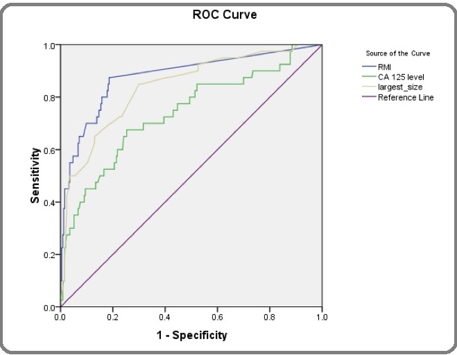
RMI score was the predictive factor with the highest sensitivity and specificity, and largest tumor size was the second best. ROC curves were drawn to evaluate useful factors for screening and to determine the optimal cutoff values (Figure 1).
Figure 1: Receiver Operating Characerisics (ROC) Curve Showing the Relattionships between Sensitivity and Specificity of Predicive Factors in Discrimination between Mature Cystic Teratoma and Malignancy Arisinng in Mature Cystic Teratoma of Ovary.

The area under the curve (AUC) for each factor was as follows: RMI score, 0.879; largest tumor size, 0.840; and CA 125, 0.739; and. This suggested that RMI score were superior to the other predictive factors for screening. Performance validation with a cut-off level of RMI > 200 showed the AUC was 0.879, with 47.5% sensitivity, 98.4% specificity, 82.6 % positive predictive value, and 92.2% negative predictive value.
Based on the ROC analysis, the sensitivity, the specificity, and the diagnostic efficiency were recalculated using the optimal cutoff values, which showed a higher diagnostic efficiency than the standard cutoff values (Figure 1 and Table 4). The best cut-off of RMI score from ROC curve was 12 with 80% sensitivity and 82.2% specificity.
| Variables | Cut-off levels | Sensitivity | Specificity | PPV | NPV | AUC |
| (%) | (%) | (%) | (%) | (95%CI) | ||
| RMI | 12 | 80 | 82.2 | 41.6 | 96.3 | 0.879 (0.813 - 0.945) |
| RMI | 200 | 47.5 | 98.4 | 82.6 | 92.2 | 0.879 (0.813-0.945) |
| Tumor size | 10 cm. | 72.5 | 77.6 | 23.6 | 96.7 | 0.840 (0.770-0.909) |
| CA 125 levels | 35 U/mL | 60 | 77.1 | 29.3 | 92.4 | 0.739 (0.648-0.830) |
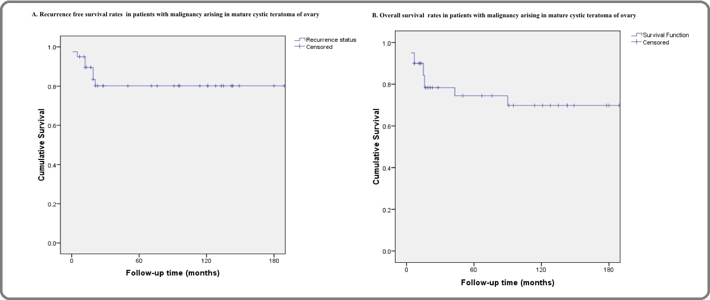
The mean and median follow-up times were 78.6 and 35.5 months, respectively. One patient (2.5%) had progressive disease after surgery and seven patients (17.5%) had recurrence. The median time interval from the diagnosis to recurrence was 12 months (range, 1-21 months). The most site of recurrent disease were pelvic recurrence (71.4%). The 5-years recurrence free survival rate was 80.2% (Figure 2A). Ten patients (25%) died of the disease during follow-up. The median time to dead was 15 months (range, 4-90 months). The overall 5-years survival rate was 74.5% (Figure 2B).
Figure 2: Survival Analysis of Patients with Malignancy Arising in Mature Cystic Teratoma of Ovary; A. Recurrence free Survival, B. Overall Survival.

Discussion
Malignant transformation arising in MCT is one of the most serious complications of MCT. Although only 0.8% to 2.4% of MCT undergo malignant transformation,the prognosis is very poor [6-8-17]. The preoperative diagnosis of malignant transformation in MCT is very difficult, and the definitive diagnosis should be provided postoperatively. By making this diagnosis prior to surgery, the most appropriate surgery can be planned and earlier consideration may be given to chemotherapy in this aggressive disease.
According to our retrospective study, we found that the incident rate of malignant transformation in our institution was 7% (40/571) which was higher than the previous reports. The explanation can be attributed to our institution being a tertiary care center where is the patients with an advanced disease or suspected malignancy are being referred from a peripheral center.
Patients with malignant transformation arising in MCT are typically postmenopausal status and may present with a rapidly enlarging tumor or symptoms suggestive of malignancy. Most of our patients also presented with palpable pelvic mass and there was consistent with previous studies [9-17]. Several literatures found that advanced age increased the risk of malignant arising in MCT whereas the present study was not showed [7-13-18]. A higher percentage (78.9%) of premenopausal women may be the explanation. Kikkawa et al. [13] and Yamanaka et al. [18] reported that a tumor diameter of greater than 9.9 cm was 86% sensitivity for malignancy in their series, and our study reported similar results. The mean tumor size of patients with malignant transform MCT in our study was 15.5 cm. compared to 8.3 cm. in benign MCT. Moreover, ROC analysis resulted in a high AUC of 0.840 which suggested that tumor size was useful in the differential diagnosis of malignant and benign MCT.
Kido et al. [19] suggested that some imaging characteristics may be helpful in the preoperative diagnosis of malignant transformation arising in MCT such as an area of the solid component with contrast enhancement, transmural extension, and irregular invasion to the peritoneal cavity. In our study, all patients had abdominopelvic ultrasonography and a multivariate analysis showed patients who had solid portions have a 5-fold risk of malignant MCT.
The usefulness of tumor markers in malignant transformation arising in MCT is not clearly understood. High concentrations of tumor markers such as SCC Ag level and CA 19-9 have been reported in patients with malignant MCT [20-22]. In our series, we could not assess these variables because of lack of data, recommendation for future clinical research to evaluate a useful tumor marker in a distinction between malignant and benign MCT include SCC Ag level, CA 19-9 levels, and CEA with a prospective design.
From the current study, the results of RMI score were within the low levels in almost all patients with benign MCT and a half of patients with malignant MCT. A low level of RMI score due to a higher percentage of stage I ovarian cancer that always no rising in CA 125 levels [23]. Another reason may be ultrasound findings showed only solid parts that also made the low level of ultrasound score. However, ROC analysis of RMI score resulted in a high AUC of 0.879 with the best cut-off of 12. Performance validation showed the sensitivity of 80% and specificity of 82.2%. These finding suggested that a high level of RMI score of greater than 12 in patients with MCT may be suspected of malignancy and guided as a tool to select these patients for referral to a gynecologic oncologist.
Due to the rarity of malignant transformation arising in MCT, there is a lack of data on the optimal treatment option. Multimodality therapy including optimal cytoreductive surgery followed by chemotherapy and/ or radiation therapy has been recommended [9-17-24- 25]. However, the appropriate adjuvant therapy for these patients has not been established. There were several previous studies supporting platinum-based chemotherapy as an effective drug for advanced ovarian cancer and SCC of the cervix [8-21-24-25]. The most of our patients were obtained optimal cytoreductive surgery and 80% of them were in stage I. All of our patients whose stage beyond IB were received adjuvant chemotherapy and the most common regimen was a combination platinum- based chemotherapy such as paclitaxel and carboplatin, paclitaxel and cisplatin, and a combination of bleomycin, etoposide, and cisplatin. Similarly, we found that most patients who receive optimal cytoreductive surgery and adjuvant treatment had an extended progression-free interval and excellent prognosis with the overall 5-year survival rate of 74.5%.
A limitation of the present study is the retrospective study design, it could be prone to a recall bias and lack of some important data such as tumor markers especially in benign MCT group. In our study, we found that serum CA 125 levels were assessed in only 253 patients with benign MCT and it may be a reason for not showing a performance validation in distinguishing between malignant benign MCT. Another limitation is a single institutional study was made, it could be limited sampling population and no validation in other population.
In conclusion, these findings demonstrate that incidence of malignancy arising in MCT was 7%. For associated risk factors, large tumor size and solid part from ultrasound finding are important factors in making a differential diagnosis of malignancy arising in MCT and MCT. Additionally, calculating RMI score might be a useful diagnostic tool to detect malignancy in this setting, and adequate staging surgery should also be considered.
Acknowledgements
This research was supported by Rajavithi research management fund.
Conflict of interest
The authors declared no conflict of interest.
References
- Malignant transformation of an ovarian mature cystic teratoma: computed tomography findings Lai Pei-Fang, Hsieh Shu-Chiang, Chien Jerry Chin-Wei, Fang Chia-Liang, Chan Wing P., Yu Chun. Archives of Gynecology and Obstetrics.2004;271(4). CrossRef
- Benign disease of the female reproductive tract. In: Berek, JS (ed.). Berek and Novak’s Gynecology, 15th ed Hillard PJA. Philadelphia: Lippincott Williams & Wilkins.;(2012):374-437.
- Ovarian Teratomas: Tumor Types and Imaging Characteristics Outwater Eric K., Siegelman Evan S., Hunt Jennifer L.. RadioGraphics.2001;21(2). CrossRef
- Squamous-cell carcinoma in mature cystic teratoma of the ovary: systematic review and analysis of published data Hackethal Andreas, Brueggmann Doerthe, Bohlmann Michael K, Franke Folker E, Tinneberg Hans-Rudolf, Münstedt Karsten. The Lancet Oncology.2008;9(12). CrossRef
- Surgical intervention for maternal ovarian torsion in pregnancy Chang Shuenn-Dhy, Yen Chih-Feng, Lo Liang-Ming, Lee Chyi-Long, Liang Ching-Chung. Taiwanese Journal of Obstetrics and Gynecology.2011;50(4). CrossRef
- Malignant transformation of ovarian mature cystic teratoma RIM S.-Y., KIM S.-M., CHOI H.-S.. International Journal of Gynecological Cancer.2006;16(1). CrossRef
- Risk factors for malignant transformation of mature cystic teratoma Park Chan-Hong, Jung Min-Hyung, Ji Yong-Il. Obstetrics & Gynecology Science.2015;58(6). CrossRef
- Malignant transformation of mature cystic teratoma of the ovary: Experience at a single institution Park Jeong-Yeol, Kim Dae-Yeon, Kim Jong-Hyeok, Kim Yong-Man, Kim Young-Tak, Nam Joo-Hyun. European Journal of Obstetrics & Gynecology and Reproductive Biology.2008;141(2). CrossRef
- Squamous Cell Carcinoma Arising in Mature Cystic Teratoma of the Ovary Hirakawa Toshio, Tsuneyoshi Masazumi, Enjoji Munetomo. The American Journal of Surgical Pathology.1989;13(5). CrossRef
- Mucoepidermoid variant of adenosquamous carcinoma arising in ovarian dermoid cyst: a case report and review of the literature KARATEKE A., GURBUZ A., KIR G., HALILOGLU B., KABACA C., DEVRANOGLU B., YAKUT Y.. International Journal of Gynecological Cancer.2006;16(S1). CrossRef
- Squamous cell carcinoma arising from dermoid cyst: Case reports and review of literature Tangjitgamol S., Manusirivithaya S., Sheanakul C., Leelahakorn S., Thawaramara T., Jesadapatarakul S.. International Journal of Gynecological Cancer.2003;13(4). CrossRef
- Clinical usefulness of serum squamous cell carcinoma antigen for early detection of squamous cell carcinoma arising in mature cystic teratoma of the ovary Miyazaki K, Tokunaga T, Katabuchi H, Ohba T, Tashiro H, Okamura H. Obstet Gynecol.1991;78(3 Pt 2):562-566.
- Diagnosis of squamous cell carcinoma arising from mature cystic teratoma of the ovary Kikkawa Fumitaka, Nawa Akihiro, Tamakoshi Koji, Ishikawa Hisatake, Kuzuya Kazuo, Suganuma Nobuhiko, Hattori Sen-ei, Furui Kenji, Kawai Michiyasu, Arii Yoshitaro. Cancer.1998;82(11). CrossRef
- Preoperative diagnosis of malignant transformation arising from mature cystic teratoma of the ovary Mori Yukiko, Nishii Hiroshi, Takabe Kazuaki, Shinozaki Hideo, Matsumoto Naoki, Suzuki Keitaro, Tanabe Hiroshi, Watanabe Akihiko, Ochiai Kazuhiko, Tanaka Tadao. Gynecologic Oncology.2003;90(2). CrossRef
- A risk of malignancy index incorporating CA 125, ultrasound and menopausal status for the accurate preoperative diagnosis of ovarian cancer JACOBS I., ORAM D., FAIRBANKS J., TURNER J., FROST C., GRUDZINSKAS J. G.. BJOG: An International Journal of Obstetrics and Gynaecology.1990;97(10). CrossRef
- Staging classification for cancer of the ovary, fallopian tube, and peritoneum Prat Jaime. International Journal of Gynecology & Obstetrics.2013;124(1). CrossRef
- Clinicopathologic study of squamous cell carcinoma of the ovary Kashimura Masamichi, Shinohara Michioki, Hirakawa Toshio, Kamura Toshiharu, Matsukuma Keita. Gynecologic Oncology.1989;34(1). CrossRef
- Preoperative diagnosis of malignant transformation in mature cystic teratoma of the ovary Yamanaka Y, Tateiwa Y, Miyamoto H, Umemoto Y, Takeuchi Y, Katayama K, et al . Eur J Gynaecol Oncol.2005;26(4):391-392.
- Dermoid cysts of the ovary with malignant transformation: MR appearance. Kido A, Togashi K, Konishi I, Kataoka M L, Koyama T, Ueda H, Fujii S, Konishi J. American Journal of Roentgenology.1999;172(2). CrossRef
- Abnormally High Values of CA 125 and CA 19-9 in Women with Benign Tumors Nagata Hiroko, Takahashi Kentaro, Yamane Yoshio, Yoshino Kazuo, Shibukawa Toshihiko, Kitao Manabu. Gynecologic and Obstetric Investigation.1989;28(3). CrossRef
- Squamous Cell Carcinoma Arising in Mature Cystic Teratoma of the Ovary Tseng Chih-Jen, Chou Hung-Hsueh, Huang Kuan-Gen, Chang Ting-Chang, Liang Ching-Chung, Lai Chyong-Huey, Soong Yung-Kuei, Hsueh Swei, Pao Chia-C.. Gynecologic Oncology.1996;63(3). CrossRef
- A Possible Genetic Factor in the Pathogenesis of Ovarian Dermoid Cysts Caspi B., Lerner-Geva L., Dahan M., Chetrit A., Modan B., Hagay Z., Appelman Z.. Gynecologic and Obstetric Investigation.2003;56(4). CrossRef
- The CA 125 tumour-associated antigen: a review of the literature Jacobs Ian, Bast Robert C.. Human Reproduction.1989;4(1). CrossRef
- Malignant Transformation Arising From Mature Cystic Teratoma of the Ovary: A Retrospective Study of 20 Cases Sakuma Michiko, Otsuki Takeo, Yoshinaga Kosuke, Utsunomiya Hiroki, Nagase Satoru, Takano Tadao, Niikura Hitoshi, Ito Kiyoshi, Otomo Keiko, Tase Toru, Watanabe Yoh, Yaegashi Nobuo. International Journal of Gynecologic Cancer.2010;20(5). CrossRef
- Squamous cell carcinoma occurring in the pelvis after total hysterectomy and bilateral salpingo-oophorectomy for an ovarian mature teratoma with malignant transformation Chen Pu, Yeh Chang-Ching, Lee Fa-Kung, Teng Sen-Wen, Chang Wen-Hsun, Wang Kuan-Chin, Wang Peng-Hui. Taiwanese Journal of Obstetrics and Gynecology.2012;51(3). CrossRef
Author Details
How to Cite
- Abstract viewed - 0 times
- PDF (FULL TEXT) downloaded - 0 times
- XML downloaded - 0 times