A Literature Review of Localized Mediastinal Mesothelioma and a Rare Case Report
Download
Abstract
Background: Primary localized mediastinal mesothelioma is a rare tumour which accounts for about 0.7% of all mesotheliomas. The mesothelial cells lining of the pericardium are suggested as the most probable cells of origin. Mediastinal pleural mesothelioma are distinctly uncommon, which has only been reported in a single case.
Case presentation: We report a case of a 57 years old man with primary mediastinal pleural mesothelioma involving middle and posterior upper mediastinum, one months following surgery, the patient is alive with good performance status. Chest plain scan and contrast-enhanced computer tomography(CT)scan showed a 43 mm×36 mm×36 mm tumor,with a CT value of about 41.5 HU, an arterial phase CT value of about 59.8HU and a venous phase CT value of about 77.5HU during the contrast-contrast-enhanced scan.Patients underwent thoracotomy.The pathological diagnosis was derived from malignant pleural mesothelioma(MPM) growing from the chest wall into the mediastinum. Conclusions Imaging manifestations of tumors have rarely been described in the literature. A short review of literature on MPM is given and summarize the imaging characteristics.
Introduction
Mediastinal mesothelioma, mainly malignant pleural mesothelioma (MPM), is rare among primary cardiac and pericardial tumors, and its incidence ranges from 0.0017% to 0.33% in case reports or collected autopsies [1]. Pericardial MPM is a rare and fatal pericardial mesothelial surface disease, accounting for 0.7% of all mesothelioma cases, with an incidence of 1 in 40 million; in 500,000 autopsies, it has an incidence of 0.0022% [2]. MPM may be present in the pleura (70 - 75%), rarely in the peritoneum (20 - 25%), pericardium (4%), or rarely elsewhere, and the incidence of localized mediastinal mesothelioma is even lower, which has only been reported in a single case [3-4]. In this report, we described the clinical presentation, imaging findings, intraoperative findings, histopathology, and immunohistochemistry of an incidentally discovered localized mesothelioma of the upper mediastinum and reviewed the relevant literature. According to the literature I reviewed, this paper is the first to summarize the imaging characteristics of all localized mediastinal mesothelioma cases.
Case report
A 57-year-old male presented with back pain without obvious inducement 9 months ago. At that time, the patient had no chest distress pain, sweating, fatigue, nausea, vomiting, abdominal pain, abdominal distension, diarrhea, etc., which could be relieved spontaneously. Later, the patient gradually developed chest distress pain, located behind the sternum, without limb cold sweating, nausea, vomiting, acid reflux, heartburn or other discomforts during the pain. About half an hour after onset, the pain could be relieved spontaneously. The patient had no obvious history of asbestos exposure. Physical examination revealed no abnormality. Coronary CTA incidentally revealed a tumor in the middle and posterior upper mediastinum (Table 1).
| Year month | 2019-8 | 2020-1 | 2020-05 | 2020-7 |
| Diagnosis and treatment process | Back pain | Coronary CTA incidentally revealed a tumor | Thoracotomy surgery | No radiotherapy and chemotherapy, The follow-up visit recovered well |
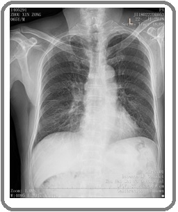
Imaging findings: An anterior chest radiograph done 8 months revealed no abnormalities (Figure 1).
Figure 1. Anteroposterior Chest Radiograph Showed no Abnormality.

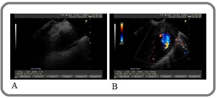
The two-dimensional B-ultrasound image showed a mild hypoechoic mass in the upper mediastinum with clear boundaries, oval shape, uneven internal echo and no change in tail echo. Color Doppler flow imaging showed no obvious blood flow signal (Figure 2).
Figure 2. Transesophageal Ultrasonography, A, Slightly Hypoechoic Mass with Clear Boundary, Oval Shape, about 12mm × 6mm in Size Can be Seen in the Upper Mediastinum Area on the Two-dimensional B-ultrasound Image. The internal echo is not uniform, and there is no change in the rear echo. B, Color doppler flow imaging showed no obvious blood flow signal.

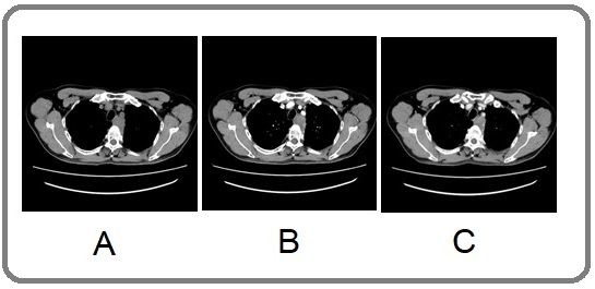
Plain scan and contrast-enhanced CT scan showed a 43 mm × 36 mm × 36 mm tumor in the middle and posterior superior mediastinum, with a CT value of about 41.5 HU, an arterial phase CT value of about 59.8HU and a venous phase CT value of about 77.5HU during the contrast- contrast-enhanced scan, with heterogeneous moderate enhancement; the tumor was adjacent to the ascending aorta, trachea, superior vena cava and right pulmonary artery. It was closely connected to the left chest wall, abutting the aortic wall, with a rough margin and indistinct demarcation from the surroundings, and no pleural or pericardial effusion was seen (Figuer 3).
Figure 3. A, is Plain CT Scan; B, is Contrast-enhanced CT Scan During the Arterial Phase; C, is Contrast-enhanced CT Scan During the Venous Phase, the Mass is Located in the Upper, Middle and Posterior mediastinum, Abutting the Pleura, with Progressive Moderate Enhancement after a Contrast-enhanced Scan.

Multidisciplinary consultation in our hospital, suspected mediastinal malignancy, and detailed systemic examination revealed no other metastatic or primary lesion, and we performed a thoracotomy. Intraoperative findings: There was a mass above the aortic arch of the posterosuperior mediastinum and in the left rear of the esophagus (about T3 - 5 level), which abutted the esophagus and aortic arch and was tightly attached to the left chest wall, and the size of the mass was about 30 mm × 25 mm × 60 mm, which was hard and had poor range of motion.
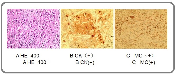
Most of the tissues were cystic wall-like, with fibrous collagenization of the capsule wall accompanied by more lymphocyte infiltration; atypical cells showed papillary, patchy, pseudo glandular, and infiltrative growth in the focal part near the surface area; tumor cells were polymorphic, mainly shuttle-shaped and oval, with abundant red staining of the cytoplasm, irregular karyotype, and visible mitoses (Figure 4).
Figure 4. A, Tumor Cells were Polymorphous, Mainly Shuttle and Oval, with Abundant Red Staining of the Cytoplasm, Irregular Karyotype, and Visible Mitoses; B, CK Immunochemical Staining was Positive; C, MK Immunochemical Staining was Positive.

Immunohistochemical results: TTF-1 (-), Napsin A (-), CK7 (partial +), P40 (-), P63 (-), CK5/6 (+), Syn (-), CgA (-), CD56 (-), Ki67 (50% +), MC (+), CK (+), Desmin (-), EMA (+), P53 (70% +), E-cad (cytoplasmic +), vimentin (+), CD5 (-), TdT (-), CD117 (-), CEA (-), PAX8 (-), CD31 (-), CD34 (-), Calretinin (good delivery, issued results -), WT1 (good delivery, issued results -). The pathological diagnosis in our hospital was derived from MPM growing from the chest wall into the mediastinum.
Follow-up and Outcomes
There has been significant progress in the treatment of malignant mesothelioma, although it remains extremely difficult to treat.
Discussion
MPM is a rare malignant tumor that is invasive and generally produces mesothelial cells such as pericardium, pleura, and peritoneum. Thoracic MPM is the most common, accounting for approximately 70% - 80% in terms of the total incidence of MPM, followed by malignant peritoneal mesothelioma [5]. MPM is characterized by a long latency period, a high degree of malignancy, and a poor prognosis. The pathogenesis of the disease is still unclear. At present, asbestos exposure is considered to be an important predisposing factor, and tuberculosis, radiation, thorotrast, etc. are also one of the predisposing factors [6-8]. Now more and more reported patients do not have a confirmed history of asbestos exposure. The main clinical symptoms are cough, chest pain, chest tightness, dyspnea, etc., and the clinical manifestations have no specificity, which brings great difficulties to its diagnosis and subsequent treatment.
Primary MPM of the pericardium accounts for less than 5% of diagnosed mesotheliomas [9]. It usually presents as multiple pericardial nodules, with or without a dominant mass, or a diffuse tumor, encasing at least a portion of the heart. There are three types of reports: extrapericardial disease (10%); localized pericardial disease (15%); diffuse nodular tumors, which can invade the heart (75%) [10-11]. Localized (solitary) pericardial mesothelioma is considered a rare variant of MPM. In 2002, J.Fernando et al. [12] reviewed only 4 cases in the literature, and S.Cao et al. [13] reported 8 cases in 2017, an increase of 4 cases compared with before. Marchevsky et al. [14] reviewed 101 cases of localized mesothelioma reported in the English literature before July 2019, which did not show an increase in the number of cases of mediastinal mesothelioma. We reviewed the English literature before May 2020, and found only 8 cases of localized (solitary) mesothelioma, with the reported cases shown in Table 2. Together with our case, there were a total of 9 cases, our case was mediastinal pleural mesothelioma, and the rest were pericardial mesothelioma. Among the 9 cases, there was only one patient with a history of predisposing factors, all of which were malignant tumors, involving 4 females and 5 males. We analyzed 3 patients with only CT scan data and 1 patient with only MRI contrast- contrast-enhanced scan data, and 1 patient with CT contrast-enhanced scan data and MRI contrast-enhanced scan data. In 4 articles, there were only CT images or MRI images,The CT image findings were not described in the article. Therefore, two senior chest attending physicians in our hospital analyzed the CT image or MRI image in the literature and our case, respectively, and obtained the image findings after discussion following listening to different views. Results showed that the image findings showed 4 cases of localized soft tissue mass, and 1 case of cystic solid lesion (cystic dominantly), after 3 months showed recurrent mass at the same site; the soft tissue density and recurrent mass was basically the same as that of muscle, 3 patients had pericardial effusion. After contrast-enhanced scan, the soft tissue CT findings showed 5 cases of mild enhancement (solid part in cystic solid lesion and recurrent mass), and 1 case of moderate enhancement; the signal after MRI T1WI weighted fat sequence enhancement was basically the same as that of muscle (Table 2).
| Case | Age/Sex | Predisposing factors of asbestos exposure | Tumor size (CM) | CT findings | MRI findings |
| 1 [14] | 27/female | NO | 4 | NO | NO |
| 2 [15] | 50/male | NO | 10 | NO | NO |
| 3 [16] | 44/female | NO | 11 | NO | Mass located above the heart, and below the aortic arch and pulmonary artery on T1WI |
| 4 [17] | 52/male | NO | 7 | A contrast-enhanced CT scan showed a 43 × 36 mm mediastinal tumor adjacent to the ascending aorta, trachea, superior vena cava, and right pulmonary artery. The mass was well encapsulated. Inhomogeneous and faint enhancement was observed in the arterial and delayed phases. No pleural or pericardial effusion was seen. | |
| 5 [18] | 59/female | NO | 6 | Pericardial effusion, enhanced CT image findings were basically the same as those in muscles. | Pericardial effusion, enhanced MRI image findings were basically the same as those in muscles. |
| 6 [19] | 68/male | NO | 3.5 (surgery) | CT scan showed a 36 × 22 mm pericardial tumor with density basically the same as muscle, pericardial effusion, moderate enhancement after contrast-enhanced scan, which was higher than muscle and lower than the aorta. | No |
| 7 [7] | 74/male | YES | 6 | Contrast-enhanced chest CT showed a right anterior mediastinal mass measuring 60 mm * 50 mm * 40 m in size, which was mainly of the cystic component. Smooth and firm cyst wall, with a uniform thickness; the mass also had soft tissue components closely related to the ascending aorta, there was no plane of separation between the tumor and the aorta, and the density was essentially the same as that of the muscle after 3 months showed recurrent mass at the same site. This mass was mainly of soft tissue attenuation without a remarkable cystic component. mild enhancement after contrast-enhanced scan. | The recurrent mass was hyperintense on both T1 and T2, Diffuse and homogeneous contrast enhancement of the mass was noted |
| 8 [12] | 76/female | NO | 3.6 | NO | NO |
| 9 | 57/male | NO | 4.3 | CT scan showed a 43 mm × 36 mm × 36 mm tumor in the middle and posterior superior mediastinum, with remarkable manifestations in the middle mediastinum, adjacent to the ascending aorta, trachea, superior vena cava and right pulmonary artery, with soft tissue density and moderate progressive homogeneous enhancement after the contrast-enhanced scan. | NO |
The CT findings of localized mediastinal mesothelioma were basically the same as those localized abdominal mesothelioma described in the article of Liu, Y.et al [15] .
In conclusions, mediastinal mesothelioma is characterized by unclear etiology, no specific manifestations, high misdiagnosis rate and poor prognosis. Pericardial mesothelioma is remarkable. Early diagnosis is beneficial to the treatment of patients. The imaging findings of localized mesothelioma in the mediastinum have not been summarized in the previous literature. This paper presents a summary. Although the number of cases is small, it has certain imaging findings. The case suggests that when the pericardium has circumscribed thickening, the mediastinal tumor is closely connected to the pleura, with the manifestation of soft tissue density, mild-moderate enhancement after contrast-enhanced scan, and pericardial effusion; the possibility of mediastinal mesothelioma should be considered.
Patient Perspective
The patient shared ideas to decrease their burden of treatment and very satisfied with the treatment.
Methods
Formal approval was granted by the Zhuhai Hospital of Guangdong Hospital of traditional Chinese Medicine.
Consent
Written informed consent was obtained form the patient for the publication of this report and any accompanying images.
Abbreviations
MPM, malignant pleural mesothelioma; CT, Computed tomography; MRI, Magnetic resonance imaging; CTCA, computer tomography Coronary artery
Availability of supporting data
The datasets used or analysed during the current study are available from the corresponding author on reasonable request.
Acknowledgements
Grateful acknowledgement is made to Dr.Ben Bishop and Dr.Mary Macalalad. who gave me considerable help by means of suggestion.
Authors’ contributions
Mengqiang xiao and Meng Zhang contributed equally to the present study. All authors read and approved the final manuscript.
Ethics approval and consent to participate
The requirement for informed consent was waived due to the retrospective nature of the study.
Competing interests
The authors declare that they have no competing interests.
References
- Surgical treatment of a primary malignant pericardial mesothelioma: case report Apicella G., Boulemden A., Citarella A., Sushma R., Szafranek A.. Acta Chirurgica Belgica.2020. CrossRef
- primary pericardial mesothelioma Eren Neyyir Tuncay, Akar A. Ruchan. Current Treatment Options in Oncology.2002;3(5). CrossRef
- Primary Pericardial Mesothelioma: A Rare Entity Godar Mohit, Liu Jianhua, Zhang Pengguo, Xia Yang, Yuan Qinghai. Case Reports in Oncological Medicine.2013;2013. CrossRef
- Localized malignant mesothelioma in the middle mediastinum: Report of a case Akamoto Shintaro, Ono Yasuhiko, Ota Kazumi, Suzaki Norikazu, Sasaki Akinori, Matsuo Yoshihiro, Hayashi Kazuhiko. Surgery Today.2008;38(7). CrossRef
- Giant malignant mesothelioma in the upper mediastinum: A case report ZHANG SHUGUANG, SONG PINGPING, ZHANG BAIJIANG. Oncology Letters.2013;6(1). CrossRef
- Mesothelioma Epidemiology, Carcinogenesis, and Pathogenesis Yang Haining, Testa Joseph R., Carbone Michele. Current Treatment Options in Oncology.2008;9(2-3). CrossRef
- Post-irradiation pericardial malignant mesothelioma: an autopsy case and review of the literature Bendek Matyas, Ferenc Miroslaw, Freudenberg Nikolaus. Cardiovascular Pathology.2010;19(6). CrossRef
- Pericardial mesothelioma following mantle field radiotherapy Velissaris T.J, et al . J Cardiovasc Surg (Torino).2001;42(3):425-427.
- Incidental localized (solitary) mediastinal malignant mesothelioma Erdogan E, Demirkazık F B, Gulsun M, Arıyurek M, Emri S, Sak S D. The British Journal of Radiology.2005;78(933). CrossRef
- Pericardial mesothelioma: An incidental intraoperative diagnosis Griffin Michael, Farmer William, Garwood Susan, Lee David. Journal of Cardiothoracic and Vascular Anesthesia.1999;13(4). CrossRef
- Primary pericardial mesothelioma unique case and literature review Sardar M.R, et al . Tex Heart Inst J.2012;39(2):261-264.
- Incidental localized (solitary) epithelial mesothelioma of the pericardium Val-Bernal J.Fernando, Figols Javier, Gómez-Román J.Javier. Cardiovascular Pathology.2002;11(3). CrossRef
- Localized malignant mesothelioma, an unusual and poorly characterized neoplasm of serosal origin: best current evidence from the literature and the International Mesothelioma Panel Marchevsky Alberto M., Khoor Andras, Walts Ann E., Nicholson Andrew G., Zhang Yu Zhi, Roggli Victor, Carney John, Roden Anja C., Tazelaar Henry D., Larsen Brandon T., LeStang Nolwenn, Chirieac Lucian R., Klebe Sonja, Tsao Ming-Sound, De Perrot Marc, Pierre Andrew, Hwang David M., Hung Yin P., Mino-Kenudson Mari, Travis William, Sauter Jennifer, Beasley Mary Beth, Galateau-Sallé Françoise. Modern Pathology.2019;33(2). CrossRef
- Malignant pericardial mesothelioma Cao S., Jin S., Cao J., Shen J., Zhang H., Meng Q., Pan B., Yu Y.. Herz.2017;43(1). CrossRef
- Localized biphasic malignant mesothelioma presenting as a giant pelvic wall mass: a rare case report and literature review Liu Yunsong, Wu Jingjun, Zhao Ying, Zhang Pengxin, Hua Zhengyu, Dong Wan, Lin Tao, Liu Ailian. BMC Medical Imaging.2020;20(1). CrossRef
- Curative resection of a well-differentiated papillary mesothelioma of the pericardium Sane, A.C , V.L. Roggli . Arch Pathol Lab Med.1995;119(3):266-267.
- Malignant mesothelioma arising from a benign mediastinal mesothelial cyst Bierhoff, E , U. Pfeifer . Gen Diagn Pathol.1996;142(1):59-62.
- Vacuolated cell mesothelioma of the pericardium resembling liposarcoma: A case report SHIMAZAKI H, AIDA S, IIZUKA Y, YOSHIZU H, TAMAJ S. Human Pathology.2000;31(6). CrossRef
- Three-year survival after surgery for primary malignant pericardial mesothelioma: report of a case Fujita Kishu, Hata Mitsumasa, Sezai Akira, Minami Kazutomo. Surgery Today.2013;44(5). CrossRef
License

This work is licensed under a Creative Commons Attribution-NonCommercial 4.0 International License.
Copyright
© Asian Pacific Journal of Cancer Care , 2021
Author Details
How to Cite
- Abstract viewed - 0 times
- PDF (FULL TEXT) downloaded - 0 times
- Xml downloaded - 0 times