Spinal Accessory Nerve Duplication – A Rare Anatomical Consternation During Neck Dissectio
Download
Abstract
Prevention of the spinal accessory nerve (SAN) is an indispensable aspect of the functional neck dissection surgery to avoid highly disabling shoulder syndrome postoperatively. This requires comprehensive knowledge of the anatomy of SAN and its variations. Rare anatomical variations like SAN duplication can result in an inadvertant injury to the SAN. We report a case of duplication of SAN, which was encountered while doing a functional neck dissection surgery for oral squamous cell carcinoma. No iatrogenic injury occurred during the surgery and neither there was any SAN dysfunction post-operatively. Meticulous dissection and consistent identification of SAN, along with vast anatomical knowledge is the key to the preservation of the nerve during the surgery. This report aims to broaden our anatomical knowledge of SAN and also discuss the clinical implications and literature pertaining to the duplication of SAN.
Introduction
In the management of head and neck cancers, neck dissection forms an essential part of the surgery. In more than a century, neck dissection surgery has evolved from the classical radical neck dissection (RND) as described by Crile [1] in 1906 to more conservative approaches like functional neck dissection (FND) and selective neck dissection (SND). While SAN was sacrificed in the RND procedure, it was Dargent and Papillon, who first proposed the preservation of SAN [2]. The preservation of SAN get its importance from the fact that iatrogenic injury to the nerve results in “Shoulder Syndrome” which is characterized by shoulder pain, impaired abduction of the shoulder and winging of scapula, thus adding to significant long term morbidity. Shoulder dysfunction has been demonstrated in up to 42.5% of patients who undergo SND [3]. Even after meticulous dissection, an iatrogenic injury to SAN can occur, especially when rare unrecognized anatomical variations like duplication are present. Knowledge of the course of SAN in the neck and its anatomical variation is imperative for the neck surgeons in order to avoid any inadvertent injury to the nerve [4]. We report a case of SAN duplication encountered during FND in a patient of oral cavity cancer.
Case Report
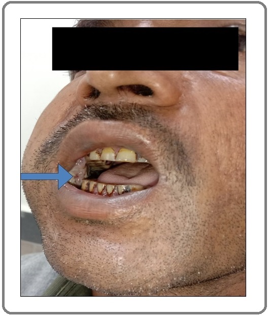
A 45-year-old male presented to the Department of Surgical Oncology with a 6-month history of a painless and gradually progressive lesion over right buccal mucosa of the oral cavity. The past history and medical history were unremarkable. The patient was a regular tobacco chewer since past 25 years. On clinical examination, an ulcero-proliferative lesion was found to involve right buccal mucosa, extending from the right canine region to the right retromolar trigone posteriorly (Figure 1).
Figure 1. Clinical Photograph Showing Lesion Over the Right Buccal Mucosa (blue arrow) with Restricted Mouth Opening.

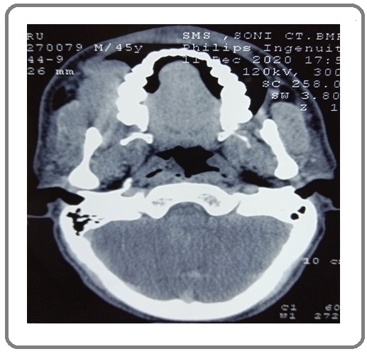
The overlying skin and underlying bone were not involved. Palpable ipsilateral cervical lymphadenopathy included level Ib, II lymph nodes. Contrast-enhanced computed tomography scan reaffirmed the findings of a 6x4cm heterogeneous lesion over the right buccal mucosa with multiple enlarged ipsilateral level Ib, II lymph nodes (Figure 2).
Figure 2. Contrast Enhanced CT Scan Showing Heterogeneous Lesion Over the Right Buccal Mucosa without any Involvement of the Underlying Bone or Overlying Skin.

Subsequently, a punch biopsy of the lesion revealed moderately differentiated squamous cell carcinoma and the patient thereafter underwent wide local excision along with modified radical neck dissection type III followed by a nasolabial flap reconstruction.
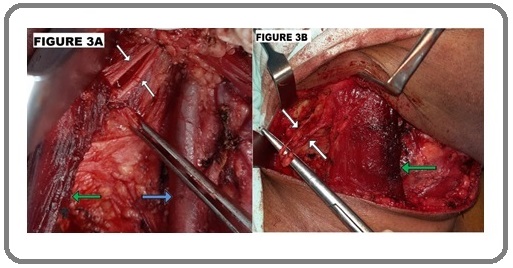
During the dissection of level IIb nodes, duplication of SAN was observed (Figure 3A), which was even more discernible during the dissection in the posterior triangle (Figure 3B).
Figure 3. Intraoperative Picture During Right Sided Modified Radical Neck Dissection Showing A) internal jugular vein (blue arrow) and duplicated spinal accessory nerve (white arrows) entering sternocleidomastoid muscle(green arrow); B) duplicated spinal accessory nerve (white arrows) traversing the posterior triangle after exiting from the lateral border of the sternocleidomastoid muscle (green arrow).

Both the nerves were seen penetrating sternocleidomastoid (SCM) muscle, then exiting SCM posteriorly before finally entering trapezius muscle. Twitching of the trapezius muscle was evident while working close to both the nerves, further elucidating the rare anatomical variation of SAN duplication. Subsequent to surgery, he recovered gradually and was discharged in a healthy condition without any shoulder dysfunction.
Discussion
SAN is the second most commonly affected peripheral nerve (18%) after median nerve (21.3%) during surgery [5]. Iatrogenic damage to the SAN is a major source of malpractice litigation [6]. Careful and meticulous dissection is the only way to preserve SAN [7] and deep knowledge of the nerve’s anatomy and its variations is thus imperative to preserve it.
After its exit from the jugular foramen, SAN descends backwards and passes deep to the posterior belly of the digastric muscle to reach the upper part of the SCM, which it innervates. It then exits SCM at its lateral border at the Erb’s point and traverses the posterior triangle obliquely downwards and backwards before it terminates in the trapezius muscle. The safest identification of the SAN is at the Erb’s point [3].
Duplication of the SAN is an extremely rare variation. SAN duplication has been reported in 2 cases out of 56 (1.8%) in a cadaveric study, one intracranially and one extracranially [8]. After an extensive review of the literature using MEDLINE, PubMed databases with a combination of following terms ‘spinal accessory nerve’; ‘dual’; ‘duplication’; ‘ anatomical variation’, we conclude that ours is the third case in the literature so far. The first case was reported in 2018 when SAN duplication was identified during neck dissection in a patient oral of cavity cancer [9]. The second case was also reported only recently, in 2020, in a patient of oral cavity cancer who underwent SND [4]. There wasn’t any iatrogenic injury to the nerve in either of the cases. Similarly, our case also had SAN duplication identified intraoperatively while doing neck dissection for oral cavity cancer.
To conclude, it is imperative to keep in mind the possible anatomical variations of SAN to avoid any inadvertent injury during neck dissection. Rare anatomical variations like duplication of SAN are mostly incidental findings encountered intraoperatively. Meticulous dissection and consistent identification of SAN along with extensive anatomical knowledge is the key to preserve the nerve during neck dissection surgery.
Acknowledgments
None
Funding
No funding was received to assist with the preparation of this manuscript.
Conflicts of interest
The authors have no conflicts of interest to declare that are relevant to the content of this article.
Consent for publication
The participant has consented to the submission of the case report to the journal.
References
- Excision of cancer of the head and neck with special breference to the plan of dissection based on one hundred and thirty-two operations. JAMA,1906;XLVII:1780-6 Crile G. https:// jamanetwork.com/journals/jama/article-abstract/369670..
- Motor complications of neck dissection: how to avoid them [in French]. Lyon Chir. 1945;40:718 Dargent M, Papillon J. .
- Spinal accessory nerve preservation in modified neck dissections: surgical and functional outcomes Popovski V., Benedetti A., Popovic-Monevska D., Grcev A., Stamatoski A., Zhivadinovik J.. Acta Otorhinolaryngologica Italica: Organo Ufficiale Della Societa Italiana Di Otorinolaringologia E Chirurgia Cervico-Facciale.2017;37(5). CrossRef
- Dual spinal accessory nerve: caution during neck dissection Danish MH, Iftikhar H, Ikram M. BMJ case reports.2020;13(6):e235487. CrossRef
- Iatrogenic Peripheral Nerve Injuries-Surgical Treatment and Outcome: 10 Years’ Experience. World Neurosurg. 2017;103:841-851.e6. 51https://www.sciencedirect.com/science/article/abs/pii/ S1878875017306071 Rasulić L, Savić A, Vitošević F, et al . . CrossRef
- Malpractice litigation after surgical injury of the spinal accessory nerve: an evidence-based analysis Morris Luc G. T., Ziff David J. S., DeLacure Mark D.. Archives of Otolaryngology--Head & Neck Surgery.2008;134(1). CrossRef
- Variations in the surface anatomy of the spinal accessory nerve in the posterior triangle Symes A, Ellis H. Surgical and radiologic anatomy: SRA.2005;27(5):404-408. CrossRef
- Variations of the accessory nerve: anatomical study including previously undocumented findings-expanding our misunderstanding of this nerve Tubbs R. Shane, Ajayi Olaide O., Fries Fabian N., Spinner Robert J., Oskouian Rod J.. British Journal of Neurosurgery.2017;31(1). CrossRef
- Spinal Accessory Nerve Duplication: A Case Report and Literature Review Papagianni Eleni, Kosmidou Panagiota, Fergadaki Sotiria, Pallantzas Athanasios, Skandalakis Panagiotis, Filippou Dimitrios. Case Reports in Otolaryngology.2018;2018. CrossRef
License

This work is licensed under a Creative Commons Attribution-NonCommercial 4.0 International License.
Copyright
© Asian Pacific Journal of Cancer Care , 2021
Author Details
How to Cite
- Abstract viewed - 0 times
- PDF (FULL TEXT) downloaded - 0 times
- XML downloaded - 0 times