A Study on Set Up Variations During Treatment, and Assessment of Adequacy of Current CTV-PTV Margins in Head and Neck Radiotherapy
Download
Abstract
Introduction: 3DCRT and IMRT demands high accuracy in patient positioning and accurate and faithful reproducibility of the treatment position right from the day of acquisition of planning scans and throughout the entire duration of radiation treatment delivery. Inadequacies in the accurate reproduction of the treatment positions during each fraction can lead to setup variations which can significantly compromise the ultimate precision of idealized treatment delivery.
Materials and methodology: We retrospectively analysed the daily setup variations in patients with head and neck malignancies who received radical or adjuvant radiotherapy from January 2018 to June 2018. A CTV-PTV margin of 0.5 cm is used at our centre. The average displacement from the reference treatment position in lateral, longitudinal and vertical directions were calculated based on CBCT shifts recorded during the entire course of treatment.
Results: 101 patients were included in the study (45.54% radical radiotherapy and 54.45% postoperative radiotherapy). The mean shift in any direction was between 0.13 cm and 0.19 cm in the study population as a whole, in radically treated patients and postoperative patients. The shift in any direction of more than 0.5 cm occurred only once or twice during the entire treatment period per patient, except one postoperative patient. The frequency of shift was more in postoperative patients. The mean shifts in the lateral and longitudinal directions were significantly more for postoperative patients (p 0.008 and 0.014 respectively).
Conclusions: The CTV to PTV expansion margin used at our institute is adequate for radically treated patients with head and neck cancers both in definitive and postoperative settings. The smaller mean shifts (<2mm) and low frequency of shifts points to the potential for reducing the current CTV to PTV expansion, which needs to be validated in larger studies.
Introduction
Head and neck cancers represent the sixth most common malignancy worldwide. It is the most common malignancy among males in India [1]. Radiotherapy plays an important role in the management of head and neck cancers. It is the standard non surgical treatment modality for locally advanced head and neck cancers. Radiotherapy is also used as adjuvant treatment with or without concurrent chemotherapy, after definitive surgery.
The goal of radiotherapy planning is to determine the optimal dose of radiation to target tissues with minimal damage to normal tissues. The modern radiotherapy treatment techniques like Three Dimensional Conformal Radiotherapy (3DCRT) and Intensity Modulated Radiotherapy (IMRT) demands high accuracy in patient positioning and accurate and faithful reproducibility of the treatment position right from the day of acquisition of planning scans and throughout the entire duration of radiation treatment delivery.
IMRT plans are usually based on pretreatment computed tomography (CT) scans that provide a snapshot of the patient’s anatomy. Hence, daily setup variations can compromise precise treatment delivery. A small margin is given around the Clinical Target Volume (CTV) to form Planning Target Volume (PTV) to allow for the set up variations that can occur during daily treatment delivery. This margin varies between institutions depending on the treatment set up at individual institutions.
In this study, we analysed the daily setup variations during radiation treatment delivery in head and neck cancer patients and the adequacy of current CTV-PTV margins used at our centre.
Materials and Methods
We retrospectively analysed the daily setup variations in patients with head and neck malignancies who received radical or adjuvant radiotherapy from 1st January 2018 to 30th June 2018. For radically treated cases, a dose of 69.3Gy to high risk volume (PTV HR), 59.4 Gy to intermediate risk volume (PTV IR) and 54 Gy to low risk volume (PTV LR) are delivered simultaneously in 33 fractions. For post operative cases, adjuvant radiotherapy dose is 60Gy to primary site and node positive regions (PTV HR) and 54 Gy to prophylactically treated regions (PTV LR) delivered simultaneously in 30 fractions. A boost dose of 6 Gy in 3 fractions is given to high risk areas with margin positivity and extranodal extension (PTV BOOST). At our centre, external beam radiotherapy for head and neck cancer patients is delivered using IMRT using volume modulated arc therapy (VMAT) with simultaneous integrated boost (SIB) technique.
A planning CT scan is taken for all patients with head and neck cancer planned for radical or adjuvant radiotherapy. Thermoplastic moulds and appropriate neck rests are used for patient immobilisation and wall mounted lasers are used to mark points on the orfit representing the reference centre. Treatment planning is done either in the Varian Eclipse or Monaco treatment planning system. A CTV-PTV margin of 0.5 cm is given to allow for the daily setup variations.
A KV Cone Beam CT scan (CBCT) with the patient in treatment position is taken on the first day of radiation treatment. After matching the bony and soft tissue anatomy in the CBCT with the reference image (planning CT scan), the final laser position representing isocenter is marked on the orfit. This absolute position in the vertical (y) lateral (x) and longitudinal (z) directions on the first day of treatment is taken as the reference or ideal position for subsequent on table verifications. CBCT is taken with patients in treatment position on the first 3 days of treatment and every two days thereafter till the completion of treatment. If the variation in any direction goes beyond 0.5 cm, and persists, a repeat planning CT scan is taken and a new plan is made.
The absolute reading of couch position that was captured during CBCT over the course of treatment was compared to the initial couch position to give an indication of the systematic and random errors. The average displacement in each direction was calculated for each patient based on CBCT shifts and is presumed to represent systematic setup error. The mean deviation in the study population as a whole, as well as in the difference in mean deviation for radical radiotherapy and adjuvant radiotherapy were analysed. The relation between different age groups and different disease stages with the mean deviations in the x, y and z directions were also analysed. The details of treatment plans and the daily shift were collected from the patient records and from the treatment planning system. The demographic data was collected from patients’ records from the hospital information system.
Statistical Analysis
Descriptive and inferential statistical methods were used for analysis. Qualitative variables were summarized using number and percentage. The normality for the set up variations in the lateral, longitudinal and vertical directions were tested using Kolmogorov Smirnov (KS) test. For the variables with normal distribution, one way Analysis of Variance (ANOVA) and independent sample t test were used to find out relation with the demographic variables. For the variable with non normal distribution, Kruskal Wallis test and Mann Whitney U test were used to test significance. The level of significance for the statistical test was fixed at 5%. Data were analyzed using SPSS statistical software, version 20.0.
Results
A total of 101 patients with head and neck cancers were treated with radical or adjuvant radiotherapy during the period. Among them, 46 (45.54%) patients received radical radiotherapy and 55(54.45%) patients received postoperative radiotherapy after radical surgery.
The median age was 62y (32-82y). Forty nine patients (48.5%) were below 60 years, 39 patients (38.6%) between 61-70 years and 13 patients (12.9%) were above the age of 70 years. In radically treated patients, the median age was 67y (48-82y) and in postoperative patients the median age was 49y (32-72y). Majority of patients were males (83/101, 82.2%) and 18 patients (17.8%) were females. The majority of patients among the postoperative group had advanced disease. The most common malignancy treated was oral cavity carcinomas followed by laryngeal carcinomas, oropharyngeal cancers and hypopharyngeal cancers. The demographic characteristics of the patients are given in Table 1.
| Total (n=101) (%) | Radical RT (n=46) (%) | Post operative RT (n=55) (%) | |
| Median age | 62y (32-82y) | 67y (48-82y) | 49y (32-72y) |
| Gender | |||
| Males | 83/101 (82.2) | 40/46 (87) | 43/55 (78.2) |
| Females | 18/101 (17.8) | 6/46 (13) | 12/55 (21.8) |
| Composite stage | |||
| Stage 1 | 9/101 (8.9) | 9/46 (19.6) | 0/55 (0) |
| Stage 2 | 14/101 (13.9) | 13/46 (28.3) | 1/55 (1.8) |
| Stage 3 | 22/101 (21.8) | 7/46 (15.2) | 15/55 (27.3) |
| Stage 4 | 56/101 (55.4) | 17/46 (37) | 39/55 (70.9) |
| Site of malignancy | |||
| Larynx | 28 (27.72) | 23 (50) | 5 (9) |
| Oropharynx | 12 (10.89) | 11 (23.9) | 1 (1.8) |
| Hypopharynx | 5 (4.95) | 4 (8.7) | 1 (1.8) |
| Nasopharynx | 3 (2.97) | 3 (6.5) | 0 (0) |
| Oral Cavity | 48 (47.52) | 2 (4.3) | 46 (83.6) |
| Maxilla | 1 (0.99) | 0 (0) | 1 (1.8) |
| Orbital Lymphoma | 2 (1.98) | 1 (2.2) | 1 (1.8) |
| Lacrimal Sac Ca | 1 (0.99) | 0 (0) | 1 (1.8) |
| Carcinoma Unknown Primary | 1 (0.99) | 1 (2.2) | 0 (0) |
Daily Set Up Variations
The mean set up variations in the lateral ( x ), longitudinal ( y ) and vertical ( z ) axes in the study population as a whole, radically treated patients and postoperative patients are given in Table 2.
| Set up variation | Total (cm) | Radical (cm) | Postoperative (cm) |
| Mean lateral | 0.17 | 0.149 | 0.182 |
| ( x ) | (min 0.04 -max 0.35) | 95% CI (0.1342,0.1641) | 95% CI (0.1628,0.2016) |
| SD 0.06 | SD 0.05 | SD 0.07 | |
| Mean longitudinal | 0.15 | 0.134 | 0.16 |
| ( y ) | (min 0.05 -max 0.36) | 95% CI (0.1146,0.1541) | 95% CI (0.1422, 0.1771) |
| SD 0.07 | SD 0.07 | SD 0.06 | |
| Mean vertical | 0.18 | 0.168 | 0.19 |
| ( z ) | (min 0.06 -max 0.57) | 95% CI (0.1428, 0.1924) | 95% CI (0.1695,0.2109) |
| SD 0.08 | SD 0.08 | SD 0.08 |
The mean deviation was 0.17 cm in the lateral direction ( x ) [0.149 for radically treated patients and 0.182 for postoperative patients]. The mean deviation in the longitudinal direction was 0.15 cm [0.134 for radically treated patients and 0.16 for postoperative patients]. The mean deviation in the vertical direction was 0.18 cm [0.168 for radically treated patients and 0.190 for postoperative patients].
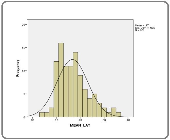
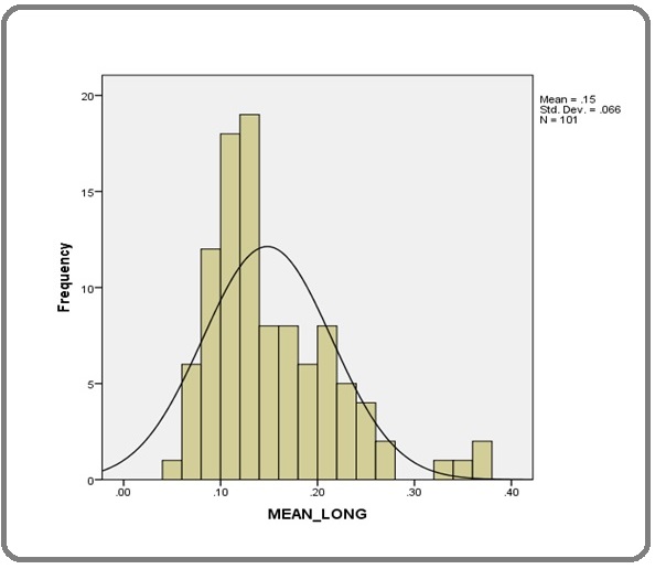
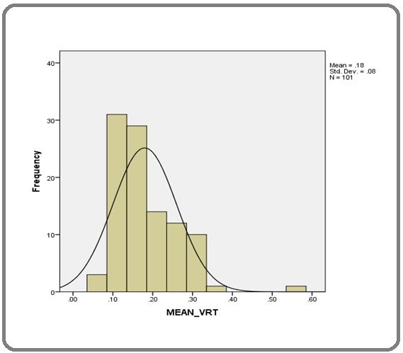
The normality for the set up variations in the lateral, longitudinal and vertical directions were tested using Kolmogorov Smirnov (KS) test. The mean lateral and the mean vertical shifts had normal distribution (KS test P-value 0.3 and 0.09 respectively), whereas mean longitudinal shift had non normal distribution (KS test P-value 0.01). The normality distributions in the mean lateral, longitudinal and vertical directions are shown in Figures 1, 2 and 3.
Figure 1. Normality of Mean LAT. Using KS Test P-value 0.327, Mean Lat has Normal Distribution.

Figure 2. Mean_ Long. Using KS Test P-value 0.010, Mean LONG has Non-normal Distribution.

Figure 3. Mean_ VRT. Using KS Test P-value 0. 093, Mean_ VRT has Normal Distribution.

Significance in mean lateral shift
There was no significant difference in mean lateral shift among different age groups (p 0.258) or between males and females (p 0.666). However, the difference was statistically significant between radical vs postoperative cases (0.15 cm vs 0.18 cm, p 0.008). A statistically significant difference in mean lateral shift was also noted among different composite stage groups (p 0.019).
Significance in mean longitudinal shift
There was no statistically significant difference in the mean longitudinal shift among different age groups (p 0.991) and between both genders (p 0.116). The difference was also not significant among different composite stage groups (p 0.407). But, there was a statistically significant difference in mean longitudinal shift between radical and postoperative cases (0.13 cm vs 0.16 cm, p 0.014).
Significance in mean vertical shift
There was no statistically significant difference in the mean longitudinal shift among different age groups (p 0.209), between both genders (p 0.417), different composite stage groups (p 0.360) as well as between radical vs postoperative cases (p 0.160).
Frequency of shifts more than 5mm
It was observed that the frequency of shifts > 5mm was more in the vertical direction (z). Out of the 101 patients, 11 (10.9%) patients had shift in the lateral direction, 8 (7.9%) patients had shift in the longitudinal direction and 24 (23.8%) patients had shift in the vertical direction.
The frequency of shifts in any direction was more in the postoperative group compared to radically treated group (14.5% vs 6.5% in the lateral direction, 9.1% vs 6.5% in the longitudinal direction and 27.3% vs 19.6% in the vertical direction for post operative patients and radically treated patients respectively). However, shifts >5 mm in any direction occurred only once or twice during the course of their treatment, except one postoperative patient who had shift >5 mm- in vertical direction three times during the treatment period. The frequency of shifts >5 mm are shown in Table 3.
| Shift >5mm | Total n (%) | Radical n (%) | Postoperative n (%) |
| Lateral | 11/101 (10.9) | 3/46 (6.5 ) | 8/55 (14.5 ) |
| Longitudinal | 8/101 (7.9) | 3/46 (6.5) | 5/55 (9.1) |
| Vertical | 24/101 (23.8) | 9/46 (19.6) | 15/55 (27.3) |
Discussion
Daily set up variations during radiation treatment may occur due to the tumour or organ motion, reduction in the tumour size or variations in patient weight or due to machine related factors. The PTV is defined to select the appropriate beam sizes and beam arrangements to ensure that the prescribed dose is actually absorbed in the CTV [2]. PTV accounts for the daily set up variations that may occur during the treatment period.
In head and neck cancers, the use of radiotherapy treatment techniques such as IMRT and IGRT has significantly improved outcome both in curative and adjuvant RT treatment. With these techniques, sharp dose gradients can be achieved with a dose distribution that tightly conforms to targets while reducing high dose to normal structures. Hence, these techniques require greater accuracy in treatment planning and daily setup during the course of radiation delivery to reduce uncertainty and setup errors.
The impact of daily setup variations on head-and-neck IMRT was studied by Hong et al [3]. They demonstrated that daily set up errors could result in significant ‘‘cold spots’’ and underdosing 1% of the tumor subvolume by 20% could lead to a loss of 11% in expected tumor control. Siberes et al [4] studied the effect of patient setup errors on simultaneously integrated boost head and neck IMRT treatment plans and noted that dose deviations upto 3% to 5% could result from random and systematic setup errors. In head and neck radiotherapy, PTV volumes are usually planned by adding 0.5cm to the CTV. Formulas including those reported by van Herk [5] and Stroom [6] are used to calculate CTV-PTV margins based on systematic and random errors reported by individual institutions. CTV to PTV margins range from 3mm to 5mm according to the immobilisation devices used and depending on the machine parameters and hence vary from institution to institution. Compared with the other tumor sites, the organ motion is insignificant in head and neck cancer patients.
The theoretical advantage of reducing CTV-to-PTV margins is to decrease dose to normal tissues, thereby improving both treatment tolerability and quality of life. Van Asselen et al [7] studied the effect of reduction of positioning margins on the dose to the parotid glands with IMRT for oropharyngeal tumors. The reduction of PTV margin from 6 mm to 3 mm, resulted in an approximately 20% reduction in normal tissue complication probability, with respect to salivary sparing.
At present, there is a lack of consensus regarding the optimal CTV-to-PTV expansion margins to be used in the treatment of head and neck cancer. Different RTOG trials exemplify this discordance. Protocol 0615 for carcinoma nasopharynx uses a minimum of 5 mm margin [8] around the CTV in all directions to define PTV and protocol 0522 [9] for carcinoma oropharynx specifically states that ‘‘a minimum margin of 3 mm can be used in all directions as long as an institution implements a study to define the appropriate magnitude of the uncertain components of the PTV’’.
The safety of PTV reduction of less than 5 mm has been demonstrated in study by Allen et al [10]. In the setting of daily IGRT, reduction of PTV margin to 3mm appeared to be adequate and did not increase local-regional failures among patients treated with IMRT for head and neck cancer. A CTV- PTV expansion margin of 3–5 mm margins may be considered appropriate only where daily IGRT [11, 12], or alternating day IGRT [13] is routine practice.
The optimal CTV-to-PTV margin depends on the method and frequency of verification imaging used in treatment. Study by Humphreys et al [14] have shown that a minimum of 5-mm margin is required when displacements are measured using bony landmarks from orthogonal plane films. However, with the increasing use of IGRT, a reduction in PTV margins may be acceptable [15].
Whether less-than-daily IGRT can serve as a viable surrogate for daily IGRT remains to be determined and is an area of active investigation. Den et al [16] in their study of 28 patients treated with IMRT for head and neck cancer noted that in the absence of daily IGRT, in order to ensure 95% of the prescribed treatment dose is delivered to 90% of the CTV, a CTV-to-PTV margins of 3.9, 4.1, and 4.9 mm are minimally required in the medial-lateral, supero-inferior, and anteroposterior dimensions respectively. An evaluation of image guidance protocols in the treatment of head and neck cancers by Zeidan et al [17] cautions against the use of less than daily temporal protocols, particularly with reduced PTV margins (i.e.,less than 5mm). In the setting of alternating day IGRT,29% and 11% of all fractions were subjected to setup errors of greater than 3 mm and 5 mm respectively. At our centre, CBCT verification is done on the first three days of starting radiation and every two days thereafter till the completion of radiation treatment. The mean shift in any direction observed in our study was less than 0.5 cm (0.13 cm - 0.19 cm) in the study population as a whole, in radically treated patients as well as postoperative patients, which denotes that our present CTV to PTV expansion margins used are adequate. The isocenter shift in any direction of more than 5 mm occurred only once or twice during the entire treatment period per patient, except one postoperative patient in whom shift occurred thrice during the treatment period. Hence, the present strategy of CBCT verifications and the CTV to PTV expansion is suitable for centres like ours with high patient load and constrained resources. However, individual institutions must derive their own CBCT verification strategy and adapt possible CTV - PTV margins.
The frequency of shift was less in radically treated patients compared to postoperative patients. Postoperative patients had higher mean lateral and mean longitudinal shifts which were statistically significant. This lesser mean shift (<2mm) and lesser frequency of shift per patient (once or twice during the treatment period) points to the potential for reducing the current CTV to PTV expansion from 5mm to 2 or 3 mm especially for patients receiving radical radiotherapy. However, the number of patients who had shift more than 5mm at least once during their treatment period, caution against the use of smaller CTV to PTV expansion. With the use of daily CBCT verifications, which have become integral part of newer machines, there is a potential for reducing the current CTV -PTV margins, thereby more of normal tissue sparing can be achieved reducing the normal tissue complication probability.
In conclusion, we conclude that the CTV to PTV expansion of 5mm is adequate for radically treated patients with head and neck cancers both in the definitive and postoperative settings. The smaller mean shifts(<2mm) and the low frequency of corrective shifts points to the potential for reducing the CTV to PTV expansion especially for patients receiving definitive radiotherapy. This needs to be validated in larger studies.
Acknowledgements
We acknowledge the continuous support of Dr,Sameer.E.P, Assistant Professor, Radiation Oncology in the compilation and finalization of the manuscript. This research did not receive any specific grant from funding agencies in the public, commercial, or not-for-profit sectors. The authors declare no conflict of interest.
References
- The Global Cancer Observatory -2020 .
- ICRU Report 50—Prescribing, Recording and Reporting Photon Beam Therapy Jones Douglas. Medical Physics.1994;21(6). CrossRef
- The impact of daily setup variations on head-and-neck intensity-modulated radiation therapy Hong Theodore S., Tomé Wolfgang A., Chappell Richard J., Chinnaiyan Prakash, Mehta Minesh P., Harari Paul M.. International Journal of Radiation Oncology, Biology, Physics.2005;61(3). CrossRef
- Effect of patient setup errors on simultaneously integrated boost head and neck IMRT treatment plans Siebers Jeffrey V., Keall Paul J., Wu Qiuwen, Williamson Jeffrey F., Schmidt-Ullrich Rupert K.. International Journal of Radiation Oncology, Biology, Physics.2005;63(2). CrossRef
- Errors and margins in radiotherapy Herk Marcel. Seminars in Radiation Oncology.2004;14(1). CrossRef
- Geometrical uncertainties, radiotherapy planning margins, and the ICRU-62 report Stroom Joep C., Heijmen Ben J. M.. Radiotherapy and Oncology: Journal of the European Society for Therapeutic Radiology and Oncology.2002;64(1). CrossRef
- The dose to the parotid glands with IMRT for oropharyngeal tumors: the effect of reduction of positioning margins Asselen Bram, Dehnad Homan, Raaijmakers Cornelis P. J., Roesink Judith M., Lagendijk Jan J. W., Terhaard Chris H. J.. Radiotherapy and Oncology: Journal of the European Society for Therapeutic Radiology and Oncology.2002;64(2). CrossRef
- Addition of bevacizumab to standard chemoradiation for locoregionally advanced nasopharyngeal carcinoma (RTOG 0615): a phase 2 multi-institutional trial Lee Nancy Y., Zhang Qiang, Pfister David G., Kim John, Garden Adam S., Mechalakos James, Hu Kenneth, Le Quynh T., Colevas A. Dimitrios, Glisson Bonnie S., Chan Anthony Tc, Ang K. Kian. The Lancet. Oncology.2012;13(2). CrossRef
- Randomized Phase III Trial of Concurrent Accelerated Radiation Plus Cisplatin With or Without Cetuximab for Stage III to IV Head and Neck Carcinoma: RTOG 0522 Ang K. Kian, Zhang Qiang, Rosenthal David I., Nguyen-Tan Phuc Felix, Sherman Eric J., Weber Randal S., Galvin James M., Bonner James A., Harris Jonathan, El-Naggar Adel K., Gillison Maura L., Jordan Richard C., Konski Andre A., Thorstad Wade L., Trotti Andy, Beitler Jonathan J., Garden Adam S., Spanos William J., Yom Sue S., Axelrod Rita S.. Journal of Clinical Oncology.2014;32(27). CrossRef
- Evaluation of the planning target volume in the treatment of head and neck cancer with intensity-modulated radiotherapy: what is the appropriate expansion margin in the setting of daily image guidance? Chen Allen M., Farwell D. Gregory, Luu Quang, Donald Paul J., Perks Julian, Purdy James A.. International Journal of Radiation Oncology, Biology, Physics.2011;81(4). CrossRef
- Evaluation of inter-fraction and intra-fraction errors during volumetric modulated arc therapy in nasopharyngeal carcinoma patients WJ Yin , Y Sun , F Chi , JL Fang , R Guo , XL Yu , et al . Radiat Oncol, 2013;8 ,article78. http://www.ro-journal.com/content/8/1/78..
- An assessment of action levels in imaging strategies in head and neck cancer using TomoTherapy. Are our margins adequate in the absence of image guidance? Houghton F., Benson R. J., Tudor G. S. J., Fairfoul J., Gemmill J., Dean J. C., Routsis D. S., Jefferies S. J., Burnet N. G.. Clinical Oncology (Royal College of Radiologists (Great Britain)).2009;21(9). CrossRef
- Comparison of daily versus nondaily image-guided radiotherapy protocols for patients treated with intensity-modulated radiotherapy for head and neck cancer Yu Yao, Michaud Anthony L., Sreeraman Radhika, Liu Tianxiao, Purdy James A., Chen Allen M.. Head & Neck.2014;36(7). CrossRef
- Assessment of a customised immobilisation system for head and neck IMRT using electronic portal imaging Humphreys Mandy, Guerrero Urbano M. Teressa, Mubata Cefas, Miles Elizabeth, Harrington Kevin J., Bidmead Margaret, Nutting Christopher M.. Radiotherapy and Oncology: Journal of the European Society for Therapeutic Radiology and Oncology.2005;77(1). CrossRef
- A comprehensive assessment by tumor site of patient setup using daily MVCT imaging from more than 3,800 helical tomotherapy treatments Schubert Leah K., Westerly David C., Tomé Wolfgang A., Mehta Minesh P., Soisson Emilie T., Mackie Thomas R., Ritter Mark A., Khuntia Deepak, Harari Paul M., Paliwal Bhudatt R.. International Journal of Radiation Oncology, Biology, Physics.2009;73(4). CrossRef
- Daily image guidance with cone-beam computed tomography for head-and-neck cancer intensity-modulated radiotherapy: a prospective study Den Robert B., Doemer Anthony, Kubicek Greg, Bednarz Greg, Galvin James M., Keane William M., Xiao Ying, Machtay Mitchell. International Journal of Radiation Oncology, Biology, Physics.2010;76(5). CrossRef
- Evaluation of image-guidance protocols in the treatment of head and neck cancers Zeidan Omar A., Langen Katja M., Meeks Sanford L., Manon Rafael R., Wagner Thomas H., Willoughby Twyla R., Jenkins D. Wayne, Kupelian Patrick A.. International Journal of Radiation Oncology, Biology, Physics.2007;67(3). CrossRef
License

This work is licensed under a Creative Commons Attribution-NonCommercial 4.0 International License.
Copyright
© Asian Pacific Journal of Cancer Care , 2022
Author Details
How to Cite
- Abstract viewed - 0 times
- PDF (FULL TEXT) downloaded - 0 times
- XML downloaded - 0 times