Dosimetric Analysis and Effect of Different Definitions of Prescription Point “A” to OAR in High Dose Rate Brachytherapy for Cervical Cancer
Download
Abstract
Purpose: Dosimetric analysis and effect of different definitions of prescription point “A” to OAR in high dose rate brachytherapy for cervical cancer.
Methods and Materials: This retrospective comparative dosimetric study is based on the data of 25 patients with histologically proven cervical carcinoma treated with HDR (high-dose-rate) brachytherapy. Patients received 21 Gy in three fractions (7.0 Gy X three fractions) to point A (AMAN, revised Manchester definition). Further, the patients were replanned with the new point A (AABS) as per the American Brachytherapy Society/ICRU 89 which is defined on CT images. The data compiled was then compared with the data observed from point A (AMAN).
Results: When AMAN normalization method was used, the mean dose to the bladder at 0.1cc, 1cc, 2cc and 5cc obtained was 1121.2±54.5, 1058.7±44.1, 875.0±38.6, 780.5±35.9, and 641.2±29.5 cGy respectively. Likewise, using the ICRU-89 point A (ABS) normalization method, the mean dose to 0.1 cc, 0.2 cc, 1 cc, 2 cc, and 5cc bladder volumes was 1178±156.1,1110.0±130.8, 921.6±111.4, 826.6±101.32, and 673.3±80.9 cGy, respectively. For the rectum, point A (MAN) normalization plans, the mean dose of 0.1 cc, 0.2 cc,1 cc, 2 cc, and 5cc rectum volumes was 680±143.5, 652.53±129.8,574.25±131.1, 530±126.9, and 452.5±121.2cGy, respectively. Likewise, using the A (ABS) plan, the mean dose of 0.1 cc, 0.2cc, 1 cc, 2 cc, and 5cc rectum volume was 707.5±148.1, 678.7±128.7, 596.2±135.6, 551.25±127.44 and 471.25±122.64 cGy respectively. The statistical mean difference of Total Reference Air Kerma rate, V100 (cc), bladder, and rectum, was found significant.
Conclusion: The present study attempts to analyze the differences in rectum and bladder dose, as well as the size of the volume enclosed for prescribed Isodose curve following ICRU 38 and ABS-ICRU 89 recommendations about intracavitary cervix cancer treated with brachytherapy. With Manchester system being more static, i.e., lesser variation with geometric changes, it is preferable in comparison to ABS system.
Introduction
Brachytherapy is used integrally as a part of treatment when carcinoma cervix patients are treated by radiation with a curative intent. It can either be used alone in case of early lesion or combined with external beam radiotherapy in advanced lesions [1-3]. Various systems are devised for dose specification for the treatment of the cervix: the two most typically used are the Manchester system and the other one is ICRU system.
The Manchester system was described by Tod and Meredith in 1938 which was later modified in 1958. It’s characterized by doses to four points: A, B, bladder and rectum. The duration of the implant relies on the dose rate at point A, which is marked 2 cm superior to the cervical os and 2 cm lateral to the cervical canal [4]. Point B is defined 3 cm laterally to point A if the central canal isn’t displaced. With the displacement of tandem with the central canal, point A moves with the canal, but point B remains fixed at 5 cm from the midline.
The revised Manchester system defined point A as the point 2cm superior to the flange and 2 cm lateral from the center of the tandem. The idea of finding point A was proposed from the bottom of the tandem loaded with radioactive sources or tandem flange since the ovoid surface wasn’t visible on plain radiographs. It includes dose specification at point A for absorbed-dose prescription, bladder and rectum dosimetry to limit dose to the organ at risk (OAR), and vaginal packing for positioning of the applicator and to spare the OAR to displace it away from the applicator. The Manchester system is still used predominantly in intracavitary brachytherapy (ICBT) in many centres [5].
There were various limitations for outlining point A because it firstly relates to the position of the sources instead of a specific anatomic structure. Secondly, dose to point A is extremely sensitive to the position of the ovoid sources relative to the tandem sources therefore can’t be the determining factor in deciding on implant duration. Thirdly counting on the dimension of the cervix, point A may lie inside or outside the tumour [6, 7].
The commission recommended a system within which the volume of tissue treated to a particular dose and also the reference air kerma (in micrograys [μGy] at 1 meter) are specified. When point A is employed as a reference point for dosing and reporting, attention must also be given to the 3D isodose distributions surrounding the entire implant.
ICRU 89 report provides recommendations on prescribing, recording, and reporting brachytherapy that specializes in volumetric imaging in cervical carcinoma brachytherapy. By using dose volume histogram ICRU 89 report combines prescribing, recording, and reporting for a simpler method of radiotherapy (planar X-ray images with a dose prescription at point A) with a sophisticated method (MR and CT images with dose prescription at D90%).
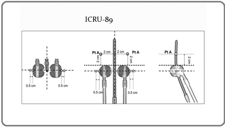
ICRU 89 and ABS recommendations: “For tandem and ovoids, connect a line through the centre of each ovoid. From the point on the tandem where this line intersects, extend superiorly along the tandem a distance equal to the radius of the ovoids” [8] (Figure 1).
Figure 1. Point A Defined by International Commission on Radiation Units and Measurements (ICRU) 89 Report.
Objective
To study the dosimetric impact of point A on the doses of the following OARs: Bladder, Rectum and Sigmoid while defined by the modified Manchester System and ICRU 89 Recommendations.
Materials and Methods
Study Population
This was a retrospective analysis of patients who received curative radiotherapy with or without concurrent cisplatin chemotherapy for cervical cancer. Patients who had Ca cervix with stages ranging from FIGO [9, 10] IB to IVA were included in this study. All patients were treated by external beam radiotherapy followed by Intracavitary brachytherapy.
Radiotherapy
EBRT was given to all patients via teletherapy cobalt machine using 2 field anterior-posterior/ posterior- anterior treatment. All patients received a dose of 50 Gy in 25 fractions. The brachytherapy was commenced after completion of EBRT. ICBT fractions were given one week apart. To calculate the dose from combined EBRT and ICBT, it was assumed that the whole volume treated with brachytherapy (including the OARs) received 100% of the prescribed EBRT dose.
Brachytherapy applicator
In ICBT, a Fletcher Suit applicator consisting of tandem and ovoids was used. It consisted of a set of two ovoids of different diameters, i.e., 20 mm and 15 mm along with the set of intrauterine tandem having different angles, i.e., 15°, 20°. The cervical stopper was used to decide the length of the intrauterine tandem.
Intracavitary insertion
The patients were well informed about the procedure and informed consent was taken. Each application was performed in the lithotomy position under LA (local anaesthesia). For identification of the ICRU bladder reference point, Foley's catheter was inserted into the bladder, and 7 cc of radio-opaque contrast was injected into the balloon. A gynaecological examination was performed, and that length of the uterine cavity was determined using a uterine sound. The applicators used were Fletcher Suit applicator.
Povidone iodine-soaked gauze was then densely packed into the vagina so that the applicator position was fixed and bladder and rectum are displaced away from the vaginal applicators. Some cotton ribbon was tied around the patients’ pelvis and then united with the applicators through the legs to stabilise the applicators during patient transfers. No systematic prescription was given for bladder filling.
Brachytherapy planning
After placement of brachytherapy applicator and Foley’s catheter, 3D images were acquired from the extent of umbilicus to mid-thigh with 1 mm slice thickness on a 64-slice diagnostic CT scan machine. High risk clinical target volume (HR-CTV) and OARs which include bladder, rectum and sigmoid colon were contoured using GYN GEC-GESTRO guidelines in Oncentra Brachy, v.4.6.0 TPS [11-13].
Contouring
The bladder, rectum, and sigmoid were contoured so as to include the full thickness of the wall of each organ. The bladder contouring started from the dome of bladder up to the junction with the urethra. For the contouring of the rectum, it began from 1 cm above the anus to the sigmoid flexure. The sigmoid colon was contoured from the recto-sigmoid flexure to the point at which the sigmoid colon extended into the anterior pelvis.
Treatment Planning
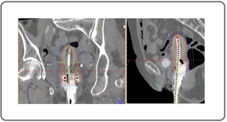
MAN In the previously treated plans, point A (A MAN ) was defined using the revised Manchester definition, i.e., 2 cm superior to the flange of the tandem and 2 cm lateral to uterine tandem. A dose of 7 Gy was prescribed to point A MAN (Figure 2) and delivered in three fractions.
Figure 2: Absolute Dose Distributions of 700 cGy Prescription to Point A as Per Revised Manchester and ICRU-89.
The optimisation of plans was done if the ICRU bladder dose exceeded 75% or rectal dose exceeded 70% of the prescribed dose. The ICRU bladder point was taken at the most posterior part of the Foley's catheter balloon. The ICRU rectal point was localised at the level of the flange on the tandem, on an anteroposterior line drawn through the tandem, 5 mm behind the posterior vaginal wall.
In addition to the ICRU points, doses to points 2.5, 5, 7.5 and 10 mm above and below these were also recorded. As the step size increases, the dose to bladder and rectum increases, therefore, the step size of 2.5 mm was used. All calculations were performed using the American Association of Physicists in Medicine Task Group – 43 (AAPM-43) algorithm on the Oncentra brachy TPS [14] as the impact of heterogeneity corrected dose calculation for nonshielded applicators is quite small in patients with cervical carcinoma. Dose volume parameters were used for reporting dose to the OAR which includes bladder, rectum, and sigmoid as per the established guidelines [11]. The parameters were estimated from the cumulative dose volume histogram for OARs.
Dose received by both point A MAN and A ABS , 2cc volume of OARs (D 2CC ) and minimum dose received by 90% and 100% of HR-CTV, i.e., D 90 and D 10C were recorded in both plans. Treatment volumes enclosed by 100% isodose line (V 100), total reference air kerma (TRAK), and geometric shifts between point A MAN and A ABS were also reported.
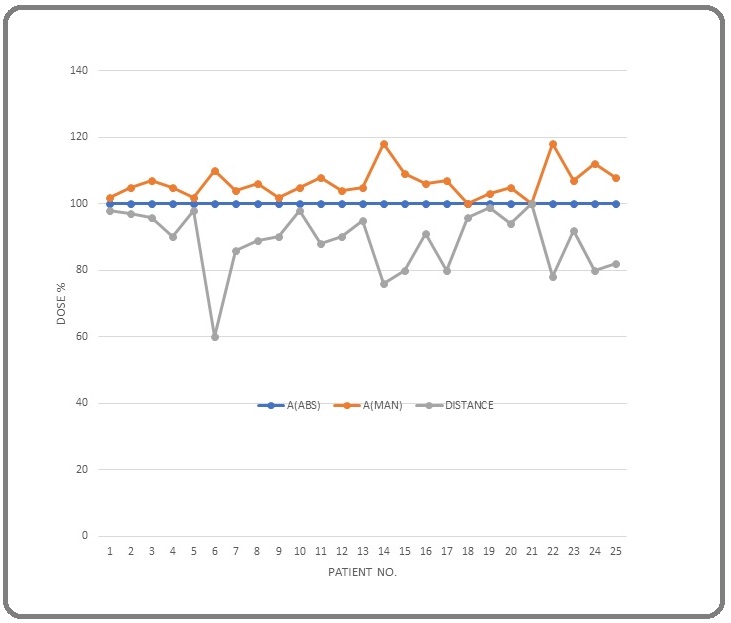
For comparison of the bladder, rectum and sigmoid doses between both the plans, mean dose of D0.1cc, D1cc and D2cc were recorded. For estimating the dose variation, difference was calculated between the 2 plans for bladder, rectum and sigmoid 0.1cc, 1cc and 2cc volumes. To see the variation in dose to point A MAN when it moves superior or inferior with respect to point A ABS we prescribed the 100% dose to point A ABS and noted the dose received at point A MAN (Figure 3) for all the patients The distance was taken positive and negative when point A MAN moves superior and inferior, respectively, to point A ABS .
Figure 3. Variation in Dose to Point A when 100% Dose is Prescribed at Point A (ABS) and A (MAN).
Results
The mean dose of 0.1 cc, 1 cc, and 2 cc volumes of the bladder, rectum are shown in Table 1 and 2.
| Bladder | P value | |||
| A (MAN) | A (ABS) | |||
| DB0.1cc | 1121.2±54.5 | 1178±156.1 | t= 1.718, Df= 48, P value= 0.092 | |
| DB0.2cc | 1058.7±44.1 | 1110.0±130.8 | t= 1.858, Df= 48, P value= 0.069 | |
| DB0 1cc | 875.0±38.6 | 921.6±111.4 | t= 1.976, Df= 48, P value= 0.054 | |
| DB0 2cc | 780.5±35.9 | 826.6±101.32 | t= 2.144, Df= 48, P value= 0.037 | |
| DB0 5cc | 641.2±29.5 | 673.3±80.9 | t= 1.864, Df= 48, P value= 0.068 |
| Rectum | P value | |||
| A (MAN) | A (ABS) | |||
| DB0.1cc | 680±143.5 | 707.5±148.1 | t= 0.667, Df= 48, P value= 0.508 | |
| DB0.2cc | 652.53±129.8 | 678.7±128.7 | t= 0.716, Df= 48, P value= 0.478 | |
| DB0 1cc | 574.25±131.1 | 596.2±135.6 | t= .582, Df= 48, P value= 0.563 | |
| DB0 2cc | 530±126.9 | 551.25±127.44 | t= .591, Df= 48, P value= 0.557 | |
| DB0 5cc | 452.5±121.2 | 471.25±122.64 | t= .544, Df= 48, P value= 0.589 |
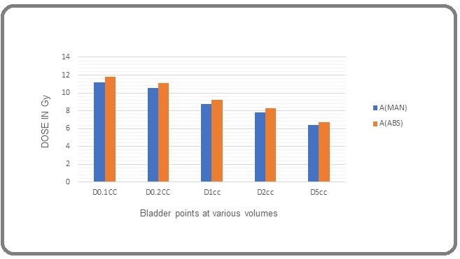
Using the A (MAN) plan normalization method, the mean dose of 0.1 cc, 0.2 cc, 1 cc, 2 cc and 5cc bladder volumes was 1121.2±54.5, 1058.7±44.1, 875.0±38.6, 780.5±35.9 and 641.2±29.5 cGy respectively. Likewise, using the ICRU-89 point A (ABS) normalization method, the mean dose of 0.1 cc, 0.2 cc, 1 cc, 2 cc and 5cc bladder volumes was 1178±156.1, 1110.0±130.8, 921.6±111.4, 826.6±101.32 and 673.3±80.9 cGy, respectively, as shown in Table 1 and 2. The mean dose difference of 0.1 cc, 1 cc, and 2 cc bladder volumes has been found significant (p <0.05) as shown in Table 1 and Figure 4.
Figure 4. Comparison of Doses at Various Bladder Points in both Manchester and ABS System.
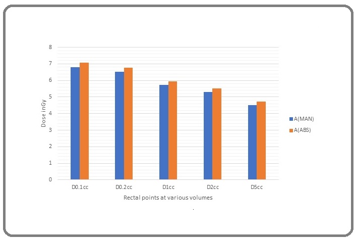
For the rectum, point A (MAN) normalization plans, the mean dose of 0.1 cc, 0.2 cc ,1 cc, 2 cc and 5cc rectum volumes was 680±143.5, 652.53±129.8, 574.25±131.1, 530±126.9 and 452.5±121.2cGy, respectively. Likewise, using the A (ABS) plan, the mean dose of 0.1 cc, 0.2cc, 1 cc, 2 cc and 5cc rectum volume was 707.5±148.1, 678.7±128.7, 596.2±135.6, 551.25±127.44 and 471.25±122.64 cGy respectively (Figure 5).
Figure 5. Comparison of Variation of Doses to Rectal Points in both Manchester and ABS System.
Discussion
Manchester point A has been used for decades as prescription point in ICBT of cervix cancer.
However, the location of this point A varies with the geometry of application. The latest ICRU-89 guidelines have recommended to use the ABS-defined point A for dose reporting.
Our findings suggest that mean point A doses of Manchester and ABS plans didn’t differ significantly.
The mean difference of point A with respect to prescription dose was 1.8% for A (MAN) and 1.2% for A (ABS). Similar results have been reported by Maurya et. al. and Anderson et. al whose values were 1.7% and 1.5% for Manchester and ABS definition respectively [15].
As shown in Table 1&2, average D2cc doses for rectum and bladder is 530cGy and 780.5cGy of prescribed dose respectively for A (MAN) and 551.25 cGy and 826.6cGy of prescribed dose respectively for A (ABS). The dose to bladder and rectum was much higher in ABS system as compared to Manchester system. The dose to bladder was found to be statistically different in both systems (p value=0.03), whereas in case of dose to rectum, differences observed was not significant in both system recommendations. (p value =.557) Similar results were derived from the study done by Howll.et.al. [16] who showed that prescribing dose to point A (ABS) leads to higher bladder and rectum dose over the prescribing dose to A (MAN), could be due to the reason that ABS point A lies superior to the above Manchester point A.
V 100 was higher for ABS plans which is similar to the observation of Anderson et.al [15] and Chang.et.al. They concluded that using ABS point A as prescription point increased the total delivered dose. The mean percentage difference in our study was 5% which was higher than finding of Anderson et. al who repored the mean percentage change of 3%.
For HR-CTV mean percentage difference between D 90 and prescribed dose was lower in ABS plans, and thus dose received by 90% volume of tumour was higher in plans using ABS point A definition for prescribing dose. There was significant difference in D 90 values of A (ABS) and A (MAN) plans. This finding is different from Anderson et. al whose mean percentage difference between D 90 parameter of two definitions was not more than 2% 90 [15].
TRAK was higher in A(ABS) by an average of 3% as compared to A(MAN) plans. This matches with Kim et. al. who showed that plans normalized to ABS point A had higher TRAK and thus prescribing to this point increased the total dose up to 2%.
The mean distance of A (ABS) point from A (MAN) point was found to be 0.6cm in our study. Zhang et.al [17] and Anderson et. al [15] calculated this shift to be 0.9cm and 0.6cm respectively.
In conclusion, the present study attempts to analyze the differences in rectum and bladder dose, as well as the size of the volume enclosed for prescribed isodose curve following ICRU 38 and ABS-ICRU 89 recommendations for brachytherapy tretment of cervical cancer. For the prescription of the treatment plans, the Manchester points (A points) were used according to ICRU 38 and the points recommended by ABS and ICRU 89. The study revealed that doses to both the points may be recorded during the transition period. However, the Manchester point Acan be preferred over the ABS point A as it conforms better with the desired outcome, is less susceptible to dose variations with geometrical changes and found to be more static.
References
- Concurrent weekly cisplatin plus external beam radiotherapy and high-dose rate brachytherapy for advanced cervical cancer: a control cohort comparison with radiation alone on treatment outcome and complications Chen Shang-Wen, Liang Ji-An, Hung Yao-Ching, Yeh Lian-Shung, Chang Wei-Chun, Lin Wu-Chou, Yang Shih-Neng, Lin Fang-Jen. International Journal of Radiation Oncology, Biology, Physics.2006;66(5). CrossRef
- Concurrent chemoradiotherapy vs. radiotherapy alone in locally advanced cervix cancer: A systematic review and meta-analysis Datta Niloy Ranjan, Stutz Emanuel, Liu Michael, Rogers Susanne, Klingbiel Dirk, Siebenhüner Alexander, Singh Shalini, Bodis Stephan. Gynecologic Oncology.2017;145(2). CrossRef
- Chemotherapy and targeted therapy in the management of cervical cancer Kumar Lalit, Harish P., Malik Prabhat S., Khurana S.. Current Problems in Cancer.2018;42(2). CrossRef
- A dosage system for use in the treatment of cancer of the uterine cervix Tod MC, Meredith WJ . Br J Radiol 1938; 11:809e824..
- Treatment of cancer of the cervix uteri, a revised Manchester method Tod M., Meredith W. J.. The British Journal of Radiology.1953;26(305). CrossRef
- Definitive radiotherapy based on HDR brachytherapy with iridium 192 in uterine cervix carcinoma: report on the Vienna University Hospital findings (1993-1997) compared to the preceding period in the context of ICRU 38 recommendations Pötter R., Knocke T. H., Fellner C., Baldass M., Reinthaller A., Kucera H.. Cancer Radiotherapie: Journal De La Societe Francaise De Radiotherapie Oncologique.2000;4(2). CrossRef
- Dose prescription, specification and applicator geometry in intracavitary therapy Potish R, Gerbi B, Engler GP. Madison, WI: Medical Physics Publishing; 1995..
- ICRU Report 89 Prescribing, Recording and Reporting Brachytherapy for Cancer of the Cervix: International Commision of Radiation Unit and Measurement, vol. 13. Oxford, UK: Oxford University Press; 2013 ;: p. 133e141. CrossRef
- Revised FIGO staging for carcinoma of the vulva, cervix, and endometrium Pecorelli Sergio. International Journal of Gynaecology and Obstetrics: The Official Organ of the International Federation of Gynaecology and Obstetrics.2009;105(2). CrossRef
- Revised FIGO staging for carcinoma of the cervix Pecorelli Sergio, Zigliani Lucia, Odicino Franco. International Journal of Gynaecology and Obstetrics: The Official Organ of the International Federation of Gynaecology and Obstetrics.2009;105(2). CrossRef
- American Brachytherapy Society consensus guidelines for locally advanced carcinoma of the cervix. Part I: general principles Viswanathan Akila N., Thomadsen Bruce. Brachytherapy.2012;11(1). CrossRef
- Recommendations from Gynaecological (GYN) GEC-ESTRO Working Group (I): concepts and terms in 3D image based 3D treatment planning in cervix cancer brachytherapy with emphasis on MRI assessment of GTV and CTV Haie-Meder Christine, Pötter Richard, Van Limbergen Erik, Briot Edith, De Brabandere Marisol, Dimopoulos Johannes, Dumas Isabelle, Hellebust Taran Paulsen, Kirisits Christian, Lang Stefan, Muschitz Sabine, Nevinson Juliana, Nulens An, Petrow Peter, Wachter-Gerstner Natascha. Radiotherapy and Oncology: Journal of the European Society for Therapeutic Radiology and Oncology.2005;74(3). CrossRef
- Recommendations from gynaecological (GYN) GEC ESTRO working group (II): concepts and terms in 3D image-based treatment planning in cervix cancer brachytherapy-3D dose volume parameters and aspects of 3D image-based anatomy, radiation physics, radiobiology Pötter Richard, Haie-Meder Christine, Van Limbergen Erik, Barillot Isabelle, De Brabandere Marisol, Dimopoulos Johannes, Dumas Isabelle, Erickson Beth, Lang Stefan, Nulens An, Petrow Peter, Rownd Jason, Kirisits Christian. Radiotherapy and Oncology: Journal of the European Society for Therapeutic Radiology and Oncology.2006;78(1). CrossRef
- Impact of heterogeneity-corrected dose calculation using a grid-based Boltzmann solver on breast and cervix cancer brachytherapy Hofbauer Julia, Kirisits Christian, Resch Alexandra, Xu Yingjie, Sturdza Alina, Pötter Richard, Nesvacil Nicole. Journal of Contemporary Brachytherapy.2016;8(2). CrossRef
- Dosimetric impact of point A definition on high-dose-rate brachytherapy for cervical cancer: evaluations on conventional point A and MRI-guided, conformal plans Anderson James, Huang Yunfei, Kim Yusung. Journal of Contemporary Brachytherapy.2012;4(4). CrossRef
- Calculation/comparison of dose to point ‘‘H’’ according to the ABS recommendations for HDR brachytherapy for carcinoma of the cervix to the dose to point ‘‘A’’ based on the revised Manchester system Howell R, Prete J, Wiatrowski W, et al . Int J Radiat Oncol 2001; 51:330e331..
- Three-dimensional dosimetric considerations from different point A definitions in cervical cancer low-dose-rate brachytherapy Zhang Miao, Chen Ting, Kim Leonard H., Nelson Carl, Gabel Molly, Narra Venkat, Haffty Bruce, Yue Ning J.. Journal of Contemporary Brachytherapy.2013;5(4). CrossRef
License

This work is licensed under a Creative Commons Attribution-NonCommercial 4.0 International License.
Copyright
© Asian Pacific Journal of Cancer Care , 2022
Author Details
How to Cite
- Abstract viewed - 0 times
- PDF (FULL TEXT) downloaded - 0 times
- XML downloaded - 0 times