Comparative Study of Dose Volume Parameters in 2-Dimensional Radiography and 3-Dimensional Computed Tomography Based High Dose Rate Intracavitary Brachytherapy in Cervical Cancer: A Prospective Study
Download
Abstract
Background: Present study compares two high-dose-rate intracavitary brachytherapy (ICBT) planning methods using two-dimensional orthogonal radiography and three-dimensional computed tomography (3D-CT) with regard to dose to target volume and organs at risk (OAR) in carcinoma cervix.
Methodology: ICBT plans for 22-patients were compared using 2D planning and three-dimensional computed tomography (3D-CT) planning techniques. 2D treatment plans were generated using 2D-orthogonal images and dose was prescribed at Point A while 3D-CT plans were generated using 3D-CT images after contouring target volume and organs at risk. In 2D planning rectal and bladder doses were assessed as per ICRU-38 and in 3D planning, 0.1cc, 0.2cc, 0.5cc and 1cc doses of bladder and rectum were evaluated. Doses to target and organ at risks (rectum and bladder) were compared for each planning method.
Results: Mean dose received by D90, D95 and D100 was 8.05±1.59Gy, 7.19±1.43Gy and 4.79±0.93Gy respectively. ICRU bladder and rectal point doses were 5.19±1.36Gy and 5.03±0.36Gy respectively. Mean dose received by bladder D0.1cc, D0.2cc, D1cc, D2cc and D5cc was 2.38±0.80, 2.22±.75, 1.85±0.64, 1.51±0.64 and 1.29±.49 times higher than ICRU bladder reference point dose. Similarly mean dose received by rectum D0.1cc, D0.2cc, D1cc, D2cc and D5cc was 1.49±0.27, 1.43±.25, 1.25±0.23, 1.09±0.21 and 0.93±.21 times higher than ICRU rectal reference point dose.
Conclusion: This study demonstrates suboptimal target coverage and underestimation of dose to OAR by 2-dimensional radiography when actual dose estimation was done by 3-dimensional brachytherapy planning for the same brachytherapy session.
Introduction
Cervical cancer is the fourth most common malignancy worldwide, and it remains the leading cause of cancer- related deaths in women, with worldwide incidence of 18,078,957 cases and 9,555,027 deaths in 2018 out of which about 80% of cases occur in developing countries. In India 96,322 women were diagnosed with cervical cancer, and 60,078 died from the disease in 2018 [1].
External beam radiotherapy (EBRT) and brachytherapy are integral Components in curative treatment of carcinoma cervix from stage IB2 to IVA [2]. Intracavitary or interstitial brachytherapy is an essential treatment modality indicated as boost after EBRT in locally advanced carcinoma cervix [3]. Conventional 2-dimensional intracavitary brachytherapy planning (ICBT) is centered on simple orthogonal X-rays and fixed bony landmarks. For 2-dimensional ICBT planning International Commission on Radiation Units and Measurement (ICRU) has recommended a number of parameters for doses and volumes for uniformity in dose reporting which includes points A and B, representing the doses in the parametria and the pelvic wall, and the rectal and bladder points representing the organs at risk (OARs). Above points are used to determine applicator position and the dose is prescribed to a fixed reference point which is 2 cm superior and lateral to the distal end of the applicator flange (Point A), irrespective of tumor characteristics and individual patient anatomy [4]. This gives rise to inadequate target coverage for large asymmetrical tumors causing treatment failure and overdose to OARs leading to increased morbidity in small tumors [5,6]. Nowadays, radiotherapy modalities have advanced rapidly with increasing sophisticated techniques which escalate the dose to tumor and spare organ at risk (OAR). Image guided brachytherapy planning (CT or MRI based) provide better volumetric information for superior target coverage and spatial relationship between applicator and OAR which leads to improved local control and reduced late toxicity [7].
Hence, present study was conducted to compare between conventional 2-Dimensional and CT based 3-Dimensional planning of HDR intracavitary brachytherapy to ascertain the potential benefit of 3D-CT based planning in terms of gross tumor volume (GTV) coverage and doses to OAR in cervical cancer patients treated with curative approach.
Public Health Implication
In Cervical cancer patients, compared to 2D treatment planning, 3D-CT image based intracavitary brachytherapy planning visualizes tumor and adjacent organ better and provides dose escalation to GTV and spare OAR (bladder & rectum), thereby reducing late toxicities and improve quality of life.
Materials and Methods
Patient characteristics
This hospital based descriptive observational study was carried out from May 2019 to April 2020. Twenty- two histopathologically proven carcinoma cervix patients, FIGO stage IIA– IIIB were enrolled prospectively in department of Radiation Oncology at S.M.S Medical College, Jaipur. The study was approved by institutional ethical committee. All patients had hematological and biochemical parameters within normal limit and had no previous history of pelvic radiotherapy and had no co-morbidities. Disease extent was evaluated with contrast enhanced magnetic resonance imaging (MRI) whole abdomen with pelvis and chest X-ray was done to rule out metastatic disease. A written informed consent was taken from all patients.
Sample size
Sample size was calculated at ALPHA error 0.05 and power 80% assuming minimum difference of means to be detected in 2D based and 3D based treatment planning, 90% planning 0.3Gy SD 0.56Gy (as per seed article), so for study purpose 30 cases of cervical cancer was planned.
Treatment scheme
All patients were treated with EBRT either by COBALT-60 teletherapy or by linear accelerator using the four-field box technique to the pelvis with a standard EBRT dose of 50 Gy in 25 fractions for 5 weeks. Bladder was kept full during EBRT to reduce dose to the small intestine. All patients received concurrent Cisplatin 30mg/m2 on first day of every week. HDR brachytherapy with cobalt-60 brachytherapy source was delivered by Eckert and Ziegler, Bebig German made HDR unit after one week of completion of EBRT. Total ICBT prescribed was 21 Gray at 7 Gray/fraction to point A in three sessions over 2 weeks.
Brachytherapy technique
Each application was performed under regional anaesthesia in the lithotomy position. Patient was advised to empty bowel before each brachytherapy session. The length of the uterine cavity was determined using a uterine sound. A foley’s catheter was inserted into the bladder and 7cc of radio-opaque contrast urograffin mixed with water (1:2 ratio) was injected into the balloon to aid in the identification of the ICRU bladder reference point. The applicators used were IBT Bebig Fletcher Suit Delcos set central tandem and ovoids. Rectal marker made of wooden stick soaked in radio-opaque dye was placed along the posterior vaginal wall. Gauze soaked with povidine iodine solution was used to pack the vagina to fix the applicators in place and to push the bladder and rectum away as much as possible from the applicator. The applicators were further stabilized with bandage, holding applicator in position with one end tied to applicator and another end wrapped on the patient’s abdomen.
Two-dimensional planning
A dedicated C-arm X-ray unit was used to take orthogonal X-rays for brachytherapy treatment planning. Point A was 2 cm superior from the mucous membrane of the cervix and 2 cm lateral to the uterine canal. Point A1 was on right side and Point A2 was on left side of central tandem. The ICRU rectal point was identified at the level of the flange on the tandem, on an antero-posterior line drawn through the tandem, 5 mm behind the posterior vaginal wall. Two additional rectal points were also taken at 3 and 6 mm above and below the ICRU point. The maximum dose (DMax) to rectum was the highest recorded dose at one of these three points. The ICRU bladder point was identified at the most posterior part of the Foley catheter balloon. Manual optimization of the plan was done starting with standard loading pattern and dwell times; adjustments were made until an optimal plan result was reached. As much as possible, the bladder dose was kept less than 80% and the rectal dose kept less than 60%. A dose of 7 Gy was prescribed to point A.
Three-dimensional computed tomography (3D-CT) planning
For each of 22-patients after first ICBT treatment session by two-dimensional planning technique, they were shifted to CT scan based ICBT planning. Intravenous contrast was injected and images taken with tandem and ovoids in-situ. CT based planning and contouring for tumor volume and OARs was done. The rectum was contoured inferiorly from lower border of obturator foramen and superiorly upto recto-sigmoid junction. The bladder was contoured from the pubic bone (bladder neck) to the superior most aspect of the bladder (dome of bladder). Clinical target volume (CTV) was contoured for every patient, which included gross tumor volume (GTV) with adequate margins. The dose per fraction from brachytherapy was given in terms of the physical dose. The minimum dose to the highest irradiated 2 cc volume of rectum and bladder were recorded (D2cc).
Comparisons were then made between the volumetric 2cc doses of the bladder and rectum with the doses at the bladder and rectum ICRU points. The dose-volume histograms (DVHs) were calculated using HDR plus Bebig Germany brachytherapy planning system.
Comparative evaluation of 2D and 3D treatment planning was done in terms of target coverage and doses to bladder and rectum i.e. ICRU-38 bladder and rectum point dose in 2D planning and dose to 0.1cc, 1cc & 2cc of bladder and rectum in 3D planning.
Statistical Analysis
Paired t-test was applied for data analysis using IBM SPSS Statistics version 22 which was used to compare 2-dimensional and 3D -CT based ICBT treatment planning results for target coverage and dose to organ at risk.
Results
Out of 22 patients, 54.4% patients were under 60-years age. Most patients presented in FIGO IIB stage (59.09%). All patients had squamous cell carcinoma histology. Table 1 shows patient characteristics.
| Patients Characteristics | No of patients (N=22) | |
| Age | <60 | 12 (54.54) |
| >60 | 10 (45.45) | |
| FIGO Stage | IIB | 13 (59.09) |
| IIIB | 9 (40.90) | |
| Histology | SCC | 22 (100) |
| Other | 0 |
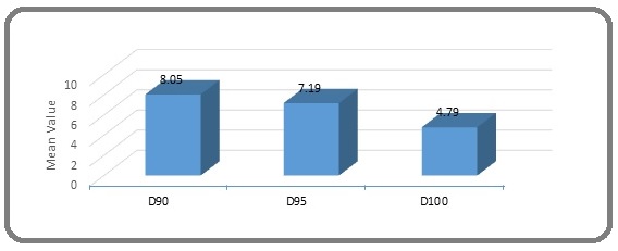
All patients were treated with EBRT followed by intracavitory brachytherapy. Mean dose prescribed at point A was 7Gy for every patient. On comparing 2D planning with 3D CT based planning, mean dose received by 90%, 95%, and 100% volume of target i.e D90, D95, D100 was 8.05±1.59, 7.19±1.43 and 4.79±0.93Gy (Table 2 and Figure 1).
| Mean dose (Gy) | Standard deviation to mean dose | Minimum dose (Gy) | Maximum dose (Gy) | |
| D90 | 8.05 | 1.59 | 5 | 10.4 |
| D95 | 7.19 | 1.43 | 4.7 | 9.3 |
| D100 | 4.79 | 0.93 | 2.7 | 5.9 |
Figure 1. Target Coverage in Three Dimensional (3D) CT Based Treatment Planning.

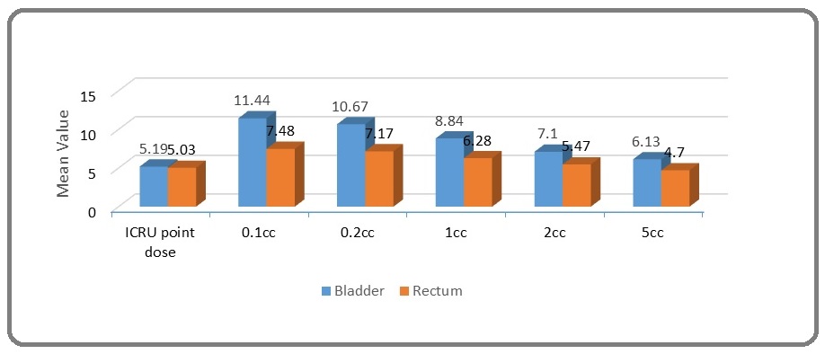
Dose received by bladder and rectum in 2D and CT based 3D planning is shown in Table 3. Paired t-test was used to compare 2D and 3D based planning results. According to 2D planning mean ICRU bladder and rectal point doses were 5.19 Gy and 5.03 Gy respectively. When 3D planning for same session was done, mean dose received by 0.1cc, 0.2cc, 1cc, 2cc and 5cc volume of bladder was 11.44 Gy, 10.67 Gy, 8.84 Gy, 7.10 Gy and 6.13 Gy respectively. Similarly mean dose received by 0.1cc, 0.2cc, 1cc, 2cc and 5cc volume of rectum was 7.48 Gy, 7.17 Gy, 6.28 Gy, 5.47 Gy & 4.70 Gy respectively (Table 3 and Figure 2).
| ICRU point dose | 0.1cc | 0.2cc | 1cc | 2cc | 5cc | |
| Bladder | 5.19±1.36 | 11.44±1.48 | 10.67±1.26 | 8.84±1.06 | 7.10±1.23 | 6.13±0.81 |
| Rectum | 5.03±0.36 | 7.48±1.40 | 7.17±1.28 | 6.28±1.23 | 5.47±1.03 | 4.70±1.08 |
Figure 2. Summary of Doses to ICRU Bladder and Rectal Reference Points in 2D Treatment Planning and Doses to 0.1cc, 1cc, 2cc, 5cc and 10cc of Bladder and Rectum using 3D DVH Parameters for OAR.

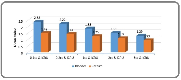
The ratio of dose received by bladder D0.1cc, D0.2cc, D1cc, D2cc and D5cc to Bladder point dose was 2.38±0.80, 2.22±.75, 1.85±0.64. 1.51±0.64 and 1.29±.49 respectively. Similarly ratio of dose received by rectum D0.1cc, D0.2cc, D1cc, D2cc and D5cc to Rectal point dose icru was 1.49±0.27, 1.43±.25, 1.25±0.23, 1.09±0.21 and 0.93±.21 respectively (Table 4 and Figure 3).
| 0.1cc/ICRU point dose | 0.2cc/ICRU point dose | 1cc/ICRU point dose | 2cc/ICRU point dose | 5cc/ICRU point dose | ||||||
| Mean | SD | Mean | SD | Mean | SD | Mean | SD | Mean | SD | |
| Bladder | 2.38 | 0.8 | 2.22 | 0.75 | 1.85 | 0.64 | 1.51 | 0.64 | 1.29 | 0.49 |
| Rectum | 1.49 | 0.27 | 1.43 | 0.25 | 1.25 | 0.23 | 1.09 | 0.21 | 0.93 | 0.21 |
Figure 3. Ratio of D0.1cc, D1cc and D2cc Doses and ICRU Point Dose for Rectum and Bladder.

When 3D planning for same session was done, difference of dose from ICRU bladder point from dose received by 0.1cc, 0.2cc, 1cc, 2cc and 5cc volume of bladder was 2.38 (95% CI 2.73, 2.02 p-value<0.001), 2.22 (95% CI 2.56, 1.89 p-value<0.001), 1.85 (95% CI 2.13, 1.56 p-value 0.0001), 1.51 (95% CI 1.79, 1.22 p-value- 0.003) and 1.29 (95% CI 1.50, 1.07 p-value-0.006).
Similarly difference of dose from ICRU rectal point to dose received by 0.1cc, 0.2cc, 1cc, 2cc and 5cc volume of rectum was 1.49 (95% CI 1.61, 1.37 p-value <0.001), 1.43 (95% CI 1.54, 1.32 p-value <0.001), 1.25 (95% CI 1.35, 1.15 p-value 0.0001), 1.09 (95% CI 1.18, 1.00 p-value 0.003), 0.93 (95% CI 1.03, 0.84 p-value 0.006) respectively (Table 5 and Figure 4).
| Bladder | P value | Rectum | p value | |
| 0.1cc & ICRU | 2.38 (95% CI 2.73, 2.02) | <0.001 | 1.49 (95% CI 1.61, 1.37 ) | p<0.001 |
| 0.2cc & ICRU | 2.22 (95% CI 2.56, 1.89) | <0.001 | 1.43 (95% CI 1.54, 1.32) | p<0.001 |
| 1cc & ICRU | 1.85 (95% CI 2.13, 1.56) | 0.0001 | 1.25 (95% CI 1.35, 1.15) | 0.0001 |
| 2cc & ICRU | 1.51 (95% CI 1.79, 1.22) | 0.003 | 1.09 (95% CI 1.18, 1.00) | 0.003 |
| 5cc & ICRU | 1.29 (95% CI 1.50, 1.07 ) | 0.006 | 0.93 (95% CI 1.03, 0.84) | 0.006 |
Figure 4. Difference between D0.1cc, D1cc and D2cc Doses and ICRU Point Dose for Rectum and Bladder.

Discussion
The conventional brachytherapy planning system based on ICRU-38 recommendations estimates dose according to reference points which are thought to represent targets and OARs. The reference points in conventional 2-D planning does not change with respect to tumor geometry, patient anatomy and OARs. An ideal conventional brachytherapy plan only ensures reproducible and comparable Point A doses between patients while it does not ensure that each patient has similar dose coverage within their tumor as each patient has different tumor extensions. Conventional 2-D planning radiation induced bladder and rectal toxicity is of utmost concern. Image based brachytherapy using CT/MRI provides precise anatomical details of target and OARs that results in better tumor control and lesser side effects. Moreover, decrease in the tumor volume after each session presents different target volumes for each brachytherapy session. In that respect, Point A and B doses offer a false sense of security since both fail to show adequate coverage of the tumor.
Present study estimated the actual doses received by target and organ at risk (rectum and bladder) when 2-D radiography-based planning was compared with 3-D ICBT volumetric planning techniques.
Gao et al found the mean target coverage of 5.4±0.9 Gy and 79.9±13.2% cc in terms of D90 and V100 respectively [8]. In our study we observed that 90%, 95% and 100% of the target volume was receiving 8.05±1.59, 7.19±1.43 and 4.79±0.93 Gy respectively, when we were prescribing 7 Gy to point A.
Kim et al observed that the mean GTV that received the prescribed dose was higher in early stage disease as compared to advanced stage and demonstrated that target coverage varied from 98.5% in stage IB1 to 59.5% in stage IIIB cervical cancer and similar results were demonstrated in other studies [9-11]. This explains poor geometry with increasing clinical stage and inadequate target coverage by the 2-D planning with consecutive sessions.
Pelloski et al observed that large tumor size itself impairs adequate GTV and CTV coverage treatment due to increased potential toxicity to bladder and rectum [12]. In our study, tumor coverage was poor for large volume tumours and dose to rectum and bladder were high compared to low volume tumours.
When CT planning of the same 2D-plan was done, we found that CTV coverage was only 88% with 7Gy dose prescription and similar results were consolidated by Fellner et who found only 83% CTV coverage with the prescribed dose of 7 Gy [11].
Present study demonstrates that dose received by 0.1cc, 0.2cc, 1cc, 2 cc and 5cc volume of bladder is 2.38±0.80, 2.22±0.75, 1.85±0.64, 1.51±0.64 and1.29±0.49 times more than ICRU bladder reference point. Similarly, dose received by 0.1cc, 0.2cc, 1cc, 2cc and 5cc volume of rectum is 1.49±0.27, 1.43±0.25, 1.25±0.23, 1.09±0.21 and 0.93±0.21 times higher than ICRU rectal reference point. Similar results were reported by previous studies because bulb of Foleys catheter does not represent the actual high dose area of bladder in 2D planning orthogonal images. Rectal point doses were similar to D2cc doses in our study [9,12,13].
Tyagi et al compared ICRU point based planning with volumetric planning in cervical cancer and concluded that bladder doses are underestimated by orthogonal film- based method but rectal doses were found similar to D 2cc doses [14]. Several other studies have also confirmed alike findings [9,12,13].
While evaluating organ-at-risk dose, the dose to 2 cc volume (D2 cc) seems to be a more meaningful reference dose, because this value is less dependent on calculation grid, is less affected by contour errors, correlates well with the maximal organ dose, and is more clinically relevant than point dose. As a result, D2 cc doses have been adopted as the criteria of OAR tolerance. The unreliability of ICRU bladder point is mainly because of the fact that its location as determined using Foley balloon does not represent the actual hottest spot in bladder.
Tharavichitkul et al reported that combination of image-based brachytherapy and intensity-modulated radiotherapy can also improve dose distribution in target volume and overdose to organs at risk [15].
Few studies have concluded that in centers where MRI based brachytherapy is not feasible due to logistics reasons, OAR and target may be contoured with CT which gives comparable results to MRI for dose volume estimation if clinical findings and baseline MRI findings are applied in contouring of CT images.
In our hospital MRI based contouring is not available so CT based planning is done using available logistics.
The limitation of our study were its small sample size due to COVID-19 outbreak and lack of MRI based brachytherapy planning as CT based planning may have overestimated the clinical target volume as compared to MRI.
In conclusion, 3-dimensional CT based brachytherapy allows precise identification and dose optimization of target volume and OAR compared to 2-dimensional based brachytherapy. The 2D conventional plan with the point A calculation relies on reference points on orthogonal films, not tumor volumes as in 3D CT plan, which may cause underestimation of tumor doses. In 2D planning, the calculation of rectum and bladder doses are made with ICRU reference points, not with bladder and rectal volumes so they may not reflect the actual organ doses. This study demonstrates suboptimal target coverage and underestimation of dose to OAR by 2-dimensional radiography based brachytherapy planning. CT based treatment planning is a reasonable substitute. 3DCT- guided brachytherapy treatment planning is recommended to overcome insufficient tumor coverage and high doses to OAR.
References
- Global cancer statistics 2018: GLOBOCAN estimates of incidence and mortality worldwide for 36 cancers in 185 countries Bray F, Ferlay J, Soerjomataram I, Siegel RL , Torre LA , Jemal A. CA: a cancer journal for clinicians.2018;68(6). CrossRef
- Figo IIIB squamous cell carcinoma of the cervix: an analysis of prognostic factors emphasizing the balance between external beam and intracavitary radiation therapy Logsdon M. D., Eifel P. J.. International Journal of Radiation Oncology, Biology, Physics.1999;43(4). CrossRef
- The American Brachytherapy Society recommendations for high-dose-rate brachytherapy for carcinoma of the cervix Nag S., Erickson B., Thomadsen B., Orton C., Demanes J. D., Petereit D.. International Journal of Radiation Oncology, Biology, Physics.2000;48(1). CrossRef
- ICRU Report no 38: Dose and volume specification for reporting intracavitary therapy in gynecology Bethesda, MD: International Commission on Radiation Units and Measurements; 1985..
- Classics in brachytherapy: Margaret Cleaves introduces gynecologic brachytherapy Aronowitz JN , Aronowitz SV , Robison RF . Brachytherapy.2007;6(4). CrossRef
- A review of recent developments in image-guided radiation therapy in cervix cancer Sadozye AH , Reed N. Current Oncology Reports.2012;14(6). CrossRef
- Proposed guidelines for image-based intracavitary brachytherapy for cervical carcinoma: report from Image-Guided Brachytherapy Working Group Nag S, Cardenes H, Chang S, Das IJ , Erickson B, Ibbott GS , Lowenstein J, Roll J, Thomadsen Br, Varia M. International Journal of Radiation Oncology, Biology, Physics.2004;60(4). CrossRef
- 3D CT-based volumetric dose assessment of 2D plans using GEC-ESTRO guidelines for cervical cancer brachytherapy Gao M, Albuquerque K, Chi A, Rusu I. Brachytherapy.2010;9(1). CrossRef
- Image-based three-dimensional treatment planning of intracavitary brachytherapy for cancer of the cervix: dose-volume histograms of the bladder, rectum, sigmoid colon, and small bowel Kim RY , Shen S, Duan J. Brachytherapy.2007;6(3). CrossRef
- Dosimetric analysis at ICRU reference points in HDR-brachytherapy of cervical carcinoma Eich H. T., Haverkamp U., Micke O., Prott F. J., Müller R. P.. Rontgenpraxis; Zeitschrift Fur Radiologische Technik.2000;53(2).
- Comparison of radiography- and computed tomography-based treatment planning in cervix cancer in brachytherapy with specific attention to some quality assurance aspects Fellner C., Pötter R., Knocke T. H., Wambersie A.. Radiotherapy and Oncology: Journal of the European Society for Therapeutic Radiology and Oncology.2001;58(1). CrossRef
- Comparison between CT-based volumetric calculations and ICRU reference-point estimates of radiation doses delivered to bladder and rectum during intracavitary radiotherapy for cervical cancer Pelloski CE , Palmer M, Chronowski GM , Jhingran A, Horton J, Eifel PJ . International Journal of Radiation Oncology, Biology, Physics.2005;62(1). CrossRef
- Comparative evaluation of two-dimensional radiography and three dimensional computed tomography based dose-volume parameters for high-dose-rate intracavitary brachytherapy of cervical cancer: a prospective study Madan R, Pathy S, Subramani V, Sharma S, Mohanti BK , Chander S, Thulkar S, Kumar L, Dadhwal V. Asian Pacific journal of cancer prevention: APJCP.2014;15(11). CrossRef
- Non isocentric film-based intracavitary brachytherapy planning in cervical cancer: a retrospective dosimetric analysis with CT planning Tyagi K, Mukundan H, Mukherjee D, Semwal M, Sarin A. Journal of Contemporary Brachytherapy.2012;4(3). CrossRef
- Preliminary results of conformal computed tomography (CT)-based intracavitary brachytherapy (ICBT) for locally advanced cervical cancer: a single institution's experience Tharavichitkul E, Mayurasakorn S, Lorvidhaya V, Sukthomya V, Wanwilairat S, Lookaew S, Pukanhaphan N, Chitapanarux I, Galalae R. Journal of Radiation Research.2011;52(5). CrossRef
License

This work is licensed under a Creative Commons Attribution-NonCommercial 4.0 International License.
Copyright
© Asian Pacific Journal of Cancer Care , 2022
Author Details
How to Cite
- Abstract viewed - 0 times
- PDF (FULL TEXT) downloaded - 0 times
- XML downloaded - 0 times