External Auditory Canal Mass: A Case Series of Squamous Cell Carcinoma
Download
Abstract
Introduction: Squamous cell carcinoma of the temporal bone is extremely rare comprising 0.2% of all tumors of head and neck. Symptoms are nonspecific and may be mistaken for other more benign conditions as otitis, cholesteatoma and polyp. Hence, this condition may present as a diagnostic challenge.
Case Discussion: We describe a case series of three patients who presented with external auditory canal (EAC) mass. The most common signs and symptoms are pruritus, decreased hearing and yellowish discharge. They were initially treated with otic antibiotics which afforded no relief. The first case is a 51 year-old female who underwent wide excision of the right ear canal and middle ear mass and right temporal craniotomy. Histopathology showed moderately differentiated squamous cell carcinoma with temporal tumor and subdural extension consistent with metastasis. The second case is a 53 year-old male who refused surgical management. He was non-compliant with work-up and follow-up and later on presented with left facial asymmetry and headache. CT scan showed a left inner and middle ear canal mass with extension to the auditory canal and protrusion into the left cerebellar hemisphere. Punch biopsy of the left EAC mass was done and pathologic report showed well-differentiated squamous cell carcinoma. Lastly, a 54 year old male who underwent Radical mastoidectomy with histopathology reports of a moderately differentiated squamous cell carcinoma.
Conclusion: Standard treatment modality is still unclear because of the rarity of this condition. However, surgical resection with negative margins followed by concurrent chemotherapy and radiation is the most commonly performed approach1. For the above-mentioned patients, concurrent chemotherapy and radiation therapy was given which showed significant treatment response.
Introduction
Squamous cell carcinoma of the temporal bone is extremely rare comprising 0.2% of all tumors of head and neck [1] with an incidence between 1 and 6 cases per million population per year [2]. The primary tumor can arise from the external auditory canal (EAC), middle ear, mastoid and petrous apex [3]. Association with chronic suppurative otitis media has been noted [2, 3]. Symptoms are nonspecific and preclude an early diagnosis. Ear pain, ear discharge and signs and symptoms that may mimic otitis, cholesteatoma and polyp are the most common early manifestations. Patients with advanced disease are seen with invasion of surrounding structures particularly the periauricular soft tissues, the parotid gland, temporomandibular joint and mastoid [4].
Standard treatment modality is still unclear because of the rarity of this condition. However, surgical resection with negative margins followed by concurrent chemotherapy and radiation is the most commonly performed approach [1].
Extensive disease, positive margins, dural and cranial nerve involvement, facial nerve paralysis and moderate to severe pain on presentation have been noted as poor prognostic factors [5]. Reported 5 year survival rates range from 40-70% but decrease to 20% in advanced disease with local recurrence as the main cause of death [6, 7].
Case Series
Three cases of squamous cell of the Temporal Bone were seen in our institution from July 2013 until August 2018 including 2 males and 1 female. The summary of the patients’ cases is shown in Table 1.
| No. | Age | Sex | Presenting Symptom | Stage at Presentation* | Treatment | Outcome |
| 1 | 51 | Female | Right ear pruritus | Stage IV T4N0M0 | Surgery→Concurrent Chemotherapy and Radiation Therapy | Progressive Disease |
| 2 | 53 | Male | Left ear discharge | Stage IV T4N0M0 | Concurrent Chemotherapy and Radiation Therapy | Progressive Disease |
| 3 | 54 | Male | Right ear purulent discharge | Stage IV T4N2bM0 | Concurrent Chemotherapy and Radiation Therapy | Stable Disease |
*Pittsburgh Staging System
Case No. 1
This is a case 51 year-old female with no known comorbid illness who presented two years prior to consult with right ear pruritus. She consulted an Ear, Nose, Throat (ENT) specialist where unrecalled otic drops was prescribed which afforded temporary relief of symptom. One year prior to consult, right ear pruritus recurred, associated with yellowish, non-foul smelling ear discharge. A small mass was noted occupying the external auditory canal. She was advised excision biopsy and histopathology showed squamous cell carcinoma. She was advised further evaluation and imaging however was lost to follow up.
Five months prior to consult, due to recurrence of the mass, she sought second opinion with another ENT specialist and Temporal Bone CT scan was done which revealed a right external auditory canal mass with osteolytic changes and extension to the middle ear, mastoid and base of the posterior cranial fossa. She was advised surgery but opted to delay the procedure.
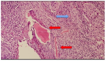
Patient came back with symptoms of right ear fullness, and tinnitus. She underwent Lateral Temporal Bone Resection, Right, with Facial Nerve Sacrifice. Intraoperatively, a fungating tumor occupies the mastoid cortex and the right external auditory canal was obliterated. Specimens were sent for Histopathology which revealed Squamous Cell Carcinoma, of the Right Ear canal, well-differentiated, with bone invasion (Figure 1).
Figure 1. Higher Magnification Shows Nests and Sheets of Atypical Polyhedral Cells with Ovoid to Irregular, Vesicular Nuclei with Prominent Nucleoli and Abundant Eosinophilic Cytoplasm (blue arrow). These atypical cells invade and destructs the adjacent bone fragment (red arrow).
Due to the extent of the tumor, a macroscopic residual disease was apparent. Patient was referred to radiation oncology and Medical oncology services.
Chest CT scan was done which showed biapical pleural thickening and subcentimeter non-calcified pleural-based nodules in both lung apices. No mass lesion or enlarged lymph nodes were noted.
On physical examination, facial asymmetry was noted on the right, with incomplete eye closure and shallow nasolabial fold. The rest of the examination was normal. Patient was advised Concurrent Chemotherapy and Radiation Therapy with Cisplatin (100 mg/m2) on days 1, 22 and 43 of External Beam Radiation Therapy 70 Gy given in 33 fractions. Patient was able to complete radiation therapy however opted not to continue chemotherapy after Cycle 1.
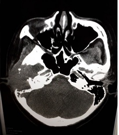
Twenty months after completion of radiotherapy, patient presented with one-month history of headache. Physical examination showed right peripheral facial palsy and decreased gross hearing, right. Repeat evaluation scans showed interval increase in size and extent of the neoplastic process involving the right external auditory canal, presently demonstrating greater degree of osseous destruction with middle ear and middle cranial fossa extension and probable skull base involvement (Figure 2).
Figure 2. Plain Temporal Bone CT Scan (Axial).
There is also temporal and parietal lobe edema with subfalcine herniation and right uncal fullness. Findings were corroborated with Cranial MRI which also showed beginning obstructive hydrocephalus.
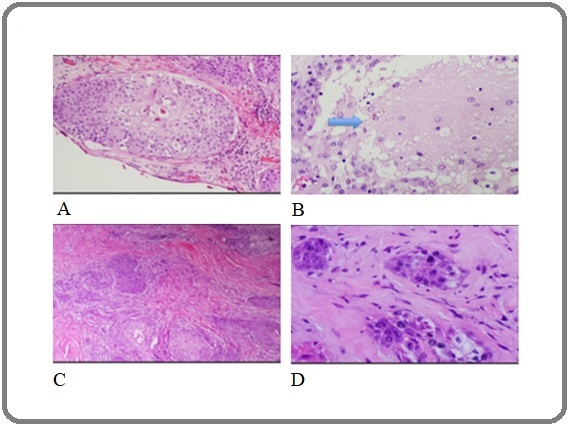
Patient underwent Right Temporal Craniotomy, Craniectomy, Excision of Tumor, Decompression, Duraplasty and Wide Excision of right external auditory canal mass, partial labyrintectomy. Histopathology report of right temporal tumor showed metastatic moderately differentiated squamous cell carcinoma with bone, dura and brain invasion. Right ear canal and middle ear mass are consistent with squamous cell carcinoma, moderately differentiated with extensive lymphovascular invasion (Figures 3A-3D).
Figure 3. A, Higher Power Magnification Shows that the Cellular Areas Show Atypical Polyhedral Cells in Nests, with Enlarged Nuclei and Abundant Eosinophilic Cytoplasm. This tumor nest invades a lymphovascular channel in the subdural section; B, In continuation with the previous slide, these tumor nests invade a portion of the brain (arrow); C, Right ear canal and middle ear mass sections show the same morphology of the tumor cells. It occurs in nests with a highly infiltrative pattern; D, Lymphovascular invasion is likewise seen in this specimen.
Patient underwent Palliative Radiation Therapy, right ear, 40 Gy divided into 20 fractions.
Case No. 2
A case of a 53 year-old male with no known comorbid illness who presented six months prior to consult with scanty watery ear discharge from the left ear with no other associated symptoms such as hearing loss, ear pain nor fullness, tinnitus, dizziness, itchiness, facial pain, nasal obstruction, blurring of vision, cough, nor colds. No consult was done nor medications taken.
Five months prior to consult, still with watery ear discharge, symptom now associated with decreased hearing and occasional tolerable pricking pain on left ear. He asked his wife to look at his ear and she noted an approximately 1x1 cm fleshy white mass protruding from the ear canal. They then decided to seek consult at a local hospital and he was given the following medications for perforated tympanic membrane, left: Ciprofloxacin 500 mg/tab 1 tab BID, Ciprofloxacin + Dexamethasone otic drops every 8 hours, and Celecoxib 400 mg/tab 1 tab BID PRN ear pain.
Four months prior to consult, patient came in for scheduled follow up. He noted no improvement of the above symptoms after taking the medications regularly. He was advised to have X-ray and/or CT scan of the ear however was not done. Patient opted to continue above medications on his own.
Two months prior to consult, after taking the medications for three months, patient noted progressive hearing loss on the left ear, hence consulted for second opinion. Mastoid Series X-ray was done showing Bilateral Mastoiditis. He was advised to undergo surgery. Audiometry was also done showing moderate to moderately severe hearing loss on the right ear and severe hearing loss at left ear. Patient continued previously prescribed medications.
One month prior to consult, wife and patient noted sudden onset of facial asymmetry, left hence consult at a tertiary hospital. He was advised to have CT scan of the temporal bone and was discharged. However, on the same day, he was recalled to be admitted. Contrast enhanced CT scan of the temporal bone showed chronic otomastoiditis with cholesteatoma formation left, mastoiditis with cholesteatoma formation right, bilateral maxillary sinusitis, hypoplastic right frontal sinus. He was given unrecalled IV antibiotics and underwent punch biopsy of the left external auditory canal mass which showed Squamous cell carcinoma, well-differentiated. Contrast enhanced MR of the brain and auditory canal showed heterogeneous soft tissue mass, left external and middle ear cavities and left mastoid, consider neoplasm, both benign and malignant, and unremarkable MRI of the brain. He was advised to undergo surgery or chemotherapy however was discharged while waiting for their decision. He opted to seek consult at our institution.
Patient is a non-smoker, occasional red wine drinker. He is fond of eating fish and vegetables and exercises (jogging) once a week for two hours. He works in an office and regularly commutes. He lives in a well-lit, well-ventilated house with no exposure to chemicals. He has no known family history of malignancy.
Physical examination was remarkable for a bluish-red mass approximately 1.3 x 0.5 cm, with serosanguinous discharge protruding from left ear canal, decreased hearing and facial palsy on the left. The rest of the examination was normal.
At the time of consult, using the Pittsburgh Staging system, he was diagnosed to have Squamous Cell Carcinoma of the External Auditory Canal, Non-keratinizing, well-differentiated, Left, Stage IV (cT4N0M0). Surgery was not feasible at this time and patient was advised to undergo concurrent chemotherapy with Cisplatin (100 mg/m2) for 3 cycles corresponding to Days 1, 22, and 43 of Radiation Therapy 70 Gy in 35 fractions. He finished 35 fractions of Radiation Therapy and only 2 cycles of Cisplatin. Patient was treated for left posterior auricular wound infection during the scheduled 2nd course of chemotherapy.
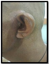
Patient tolerated the treatment well with noted significant resolution of the ear canal mass (Figure 4).
Figure 4. Left Ear. Resolution of left ear canal mass after concurrent chemotherapy and radiation therapy.
There was residual hearing loss on the left, facial asymmetry and mild numbness of the face.
During the last evaluation in February 2018, Chest CT scan showed subcentimeter pulmonary nodules in the lingula and abutting the major fissure, largest of which measures 0.3 cm adjacent to the pleura. Calcified pulmonary nodules are also seen in the apico-posterior segment of the left upper lobe and right minor fissure. There was no evidence of enlarged mediastinal and hilar lymph nodes. Of note is an incidental finding of a 2.7 x 1.7 cm enhancing soft tissue in the visualized left upper hemiabdomen. Patient was advised Upper Abdominal CT Scan for further evaluation however was lost to follow up. In July 2018, patient presented with inability to urinate, hypogastric pain with direct tenderness, and numbness of bilateral plantar aspect of the feet with difficulty standing up and eventual paralysis. Thoracic Spine MRI showed enhancing intramedullary spinal cord lesions are seen at the level of C7 to T1, the largest of which measures 1 x 1.2 x 1.4 cm with leptomeningeal enhancement. There is spinal cord edema from C5-C6 down to the level of T4 with effacement of the subarachnoid space; a similar enhancing focus with perilesional edema is also noted at the level of T7-T8 measuring 0.5 x 0.3 x 0.6 cm (Figure 5).
Figure 5. Thoracic Spine MRI (Sagittal).
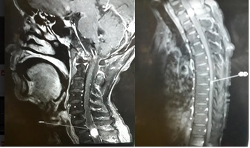
Minimal patchy degenerative fatty marrow changes are seen. There was an incidental note of enhancing lesion in the basal frontal region. Multilevel cervical spondylosis is also seen. Significant findings on physical examination are peripheral facial palsy, left; complete hearing loss, left; 0/5 motor with 0% sensory, and areflexia on bilateral lower extremities. Patient was managed as a case of Spinal Cord Compression and was started on Dexamethasone and advised Radiation Therapy. However, patient was lost to follow up.
Case No. 3
A 54 year-old male presented with one month history of purulent discharge of the right ear. No other signs and symptoms such as ear pain nor fullness, tinnitus, dizziness, itchiness, facial pain, nasal obstruction, blurring of vision, cough, nor colds. He underwent left mastoidectomy during his childhood and was functionally deaf since.
Patient is a previous smoker consuming 3 sticks per day for 30 years and stopped a month prior to diagnosis. He is not an alcoholic beverage drinker. There was also no history of malignancy among the 1st degree relatives. Patient was initially managed in another institution where he underwent Radical Mastoidectomy for Chronic Otitis Media, Right. Histopathology showed moderately differentiated Squamous Cell Carcinoma, Right mastoid. He was referred to Radiation Oncology and Medical Oncology services for further evaluation and management.
Upon consultation, he complained of headache not relieved with Tramadol. There was a note of a fungating mass at the right ear canal with ulceration at the posterior auricular area and palpable right cervical lymphadenopathy. The rest of the examination was unremarkable. Head and Neck Surgeon evaluated the lesion which was deemed unresectable.
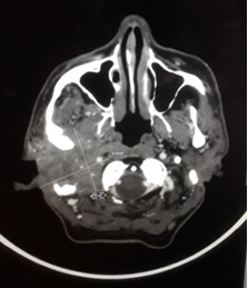
Metastatic work up was requested prior to treatment planning. CT Scan of the Brain, Neck and Temporal Bone showed a large enhancing inhomogeneous mass lesion measuring about 5.8 x 6.2 x 7.3 cm centered in the right mastoid/temporal region extending anteriorly into the right posterior temporal convexity of the brain, right temporal fossa and posteriorly into the region of the right jugular foramen/sigmoid plate, superiorly into the right middle cranial fossa down to the level of C3-C4 infiltrating the right parotid gland. There are also erosive changes of the right mastoid bone, sigmoid plate, jugular foramen, temporal fossa and floor of the right middle cranial fossa. There is encasement of the posterolateral wall of the petrous segment of the right internal carotid artery. The labyrinthine segment of the right facial nerve canal is widened with non-demonstration of rest of the facial nerve canal segment raising the possibility of infiltration. There is a large defect in the left mastoid bone, possibly from previous canal wall down radical mastoidectomy. There are enlarged lymph nodes, right, level IIA, right level IIB, inferior periparotid area and right level III. No enlarged lymph nodes on the left side (Figure 6).
Figure 6. Contrast-enhanced Temporal Bone Scan (Axial).
Patient underwent concurrent chemotherapy with Carboplatin (70 mg/m2) Days 1-4 + 5-Fluorouracil (600 mg/m2) Days 1-4 every 21 days for 3 cycles corresponding to Days 1, 22, and 43 of Intensity-Modulated Radiation therapy (IMRT) using Varian Linear accelerator 70 Gy given in 35 daily fractions with overall treatment of 50 days. Post Cycle 1 Carboplatin, there was resolution of headache and ulceration on the right posterior auricular area dried up. During the course of treatment, occasional difficulty in swallowing and erythema of the right side of the neck and face were noted. Otherwise, patient tolerated the treatment well.
On surveillance, there was no subjective complaint. Patient has improved appetite, no ear discharge nor headache. No new lesions were seen at the right aural area. Evaluation CT scan showed no discrete enhancing mass in the right mastoid/temporal region to suggest tumor recurrence. The rest of the findings were stable.
Discussion
There is no common predisposing factor for the patients in this series. Literature suggests that chronic suppurative otitis media may be a risk factor, and this was documented in one of the cases. The demographic variables show that all three patients were in their 5th decade of life during the time of diagnosis. Although there is no sex preponderance for squamous cell carcinoma of the EAC, previous reports show that patients are predominantly male [8] as was seen in our current series.
All patients presented with nonspecific signs and symptoms, the most common of which is ear discharge. Other associated symptom was headache, ear pain, ear pruritus and hearing loss. However, since other benign conditions may also present with the similar findings, diagnosis of squamous cell carcinoma was determined later on in the course. All patients have Stage IV disease at the time of diagnosis.
Due to the rarity of the disease, an adequate staging system has not been developed [9]. Currently, the University of Pittsburgh staging system is being used. This staging however is limited in determination of soft tissue extension. The modified Pittsburgh staging system classifies presence of facial paralysis as T4 tumors.
Surgery is still the cornerstone of treatment for this malignancy. Complete removal and surgical-free margins should be sought followed by radiotherapy in all cases [10]. Tiwari et al [11] described the operative technique of Subtotal Bone Resection with postoperative radiotherapy. Five-year Disease Free Survival (DFS) was 42% following this combined treatment modality. On the other hand, a recurrence rate of 67% was noted in patients with positive margins [12]. Ogawa et al [13] recommends radical radiotherapy for T1 disease and surgery with radiotherapy for T2-T3 disease. On multivariate analysis, T stage and treatment modality were significant prognostic factors. This multi-institutional retrospective review of 87 patients showed that the 5-year DFS in patients with negative surgical margins is 83%, those with positive surgical margins is 55% and those with macroscopic residual disease was 38% (P=0.007). Further studies on the role of chemotherapy are needed.
In this series, one of the patients underwent surgical treatment due to the extent of invasion, there was a residual tumor. Post-operatively, she underwent definitive chemoradiation therapy but was only able to complete the radiation therapy. The lesion progressed 20 months after treatment and Radiation therapy 40 Gy divided in 20 fractions was administered. Patient eventually succumbed to the disease.
The second patient initially refused surgery, thus, at the time of consult in our institution, the mass was already unresectable. He was compliant with the chemoradiation treatment and was able to achieve stable disease with significant regression in the size of the ear canal mass. Patient was lost to follow up and presented with Spinal Cord Compression nine months after completion of treatment. And although Squamous cell carcinoma of the temporal bone recurs locally, probable distant metastasis in this case further emphasizes the aggressive behavior of the disease and the need for close surveillance. Facial palsy at the time of diagnosis was noted to be a poor prognostic factor [4] and this was seen in this case.
The third case also has unresectable disease at the time of diagnosis. He responded well with the treatment and still has stable disease as of the last evaluation.
Early diagnosis and high index of suspicion cannot be overemphasized in this disease. Due to the aggressive behavior of Squamous cell carcinoma of the EAC, radical treatment with combined modality and vigilant surveillance and monitoring should be employed.
Acknowledgements
We are indebted to the Institutes of Pathology and Radiology for the images shown in this report.
References
- Squamous Cell Carcinoma of the External Auditory Canal in the Young: A rare case report Sandeep Yadav , Devendra Gupta , GS Yaday . 2013;19(4):196-198.
- Squamous cell carcinoma of the external auditory canal Lobo D, Llorente jl , Suárez C. Skull Base: Official Journal of North American Skull Base Society ... [et Al.].2008;18(3). CrossRef
- Temporal bone squamous cell carcinoma - Penang experience Ng SY , Pua KC , Zahirrudin Z. The Medical Journal of Malaysia.2015;70(6).
- Primary squamous cell carcinoma of the external auditory canal: surgical treatment and long-term outcomes Mazzoni A, Danesi G, Zanoletti E. Acta Otorhinolaryngologica Italica: Organo Ufficiale Della Societa Italiana Di Otorinolaringologia E Chirurgia Cervico-Facciale.2014;34(2).
- Prognostic factors in carcinoma of the external auditory canal Testa JR , Fukuda Y, Kowalski LP . Archives of Otolaryngology--Head & Neck Surgery.1997;123(7). CrossRef
- Localized carcinoma of the external ear is an unrecognized aggressive disease with a high propensity for local regional recurrence Yoon M, Chougule P, Dufresne R, Wanebo HJ . American Journal of Surgery.1992;164(6). CrossRef
- Distant metastases from ear and temporal bone cancer Sasaki CT . ORL; journal for oto-rhino-laryngology and its related specialties.2001;63(4). CrossRef
- Analysis of 95 cases of squamous cell carcinoma of the external and middle ear Yin M, Ishikawa K, Honda K, Arakawa T, Harabuchi Y, Nagabashi T, Fukuda S, et al . Auris, Nasus, Larynx.2006;33(3). CrossRef
- Squamous Cell Carcinoma of the External Auditory Canal: A Case Report. Case Reports in Otolaryngology Harry Boamah , Glenn Knight , Joseph Taylor , Kevin Palka , Billy Ballard . ;Volume 2011, Article ID 615210.
- The role of radiotherapy in treating squamous cell carcinoma of the external auditory canal, especially in early stages of disease Hashi N., Shirato H., Omatsu T., Kagei K., Nishioka T., Hashimoto S., Aoyama H., Fukuda S., Inuyama Y., Miyasaka K.. Radiotherapy and Oncology: Journal of the European Society for Therapeutic Radiology and Oncology.2000;56(2). CrossRef
- Squamous cell carcinoma of the external auditory canal and middle ear results of treatment with subtotal temporal bone resection and postoperative radiotherapy Tiwari R, Brouwer J, Quak J, Tobi H, Winters H, Mehta D. Indian Journal of Otolaryngology and Head & Neck Surgery.2002;54(3). CrossRef
- Cancer of the external auditory canal Nyrop M, Grøntved A. Archives of Otolaryngology--Head & Neck Surgery.2002;128(7). CrossRef
- Treatment and prognosis of squamous cell carcinoma of the external auditory canal and middle ear: a multi-institutional retrospective review of 87 patients Ogawa K, Nakamura K, Hatano K, Uno T, Fuwa N, Itami J, Kojya S, et al . International Journal of Radiation Oncology, Biology, Physics.2007;68(5). CrossRef
License

This work is licensed under a Creative Commons Attribution-NonCommercial 4.0 International License.
Copyright
© Asian Pacific Journal of Cancer Care , 2023
Author Details
How to Cite
- Abstract viewed - 0 times
- PDF (FULL TEXT) downloaded - 0 times
- XML downloaded - 0 times